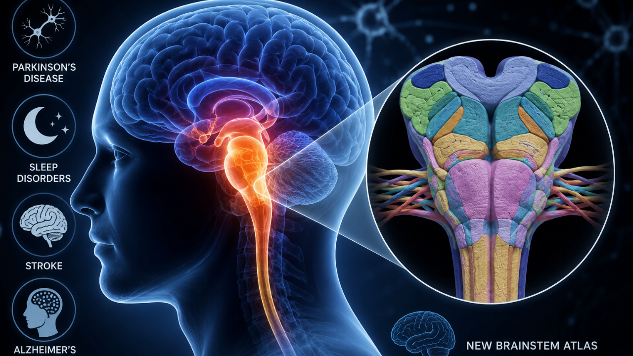
Indian researchers have created the world's most detailed three-dimensional atlas of the human brainstem. This breakthrough could significantly advance research into neurological conditions like Parkinson's disease, sleep disorders, stroke, and Alzheimer's disease. Developed by researchers at the Sudha Gopalakrishnan Brain Centre at IIT Madras, the atlas is called ANCHOR (Atlas of Neurochemical Characterization of the Human Brainstem with 3D Reconstruction).Also Read: US Senator & Trump's Close Ally Lindsey Graham Died Of Aortic Dissection: All About The Fatal Heart Emergency It offers an unprecedented view of one of the brain's most vital yet least understood regions. The atlas integrates MRI scans, microscopic tissue imaging, and neurochemical mapping to reveal the brainstem at cellular resolution across prenatal, childhood, and adult stages. About The Human Brainstem The brainstem controls all the essential functions that keep us alive, including breathing, heart rate, sleep, consciousness, body temperature, and movement. It also serves as the communication bridge between the brain and the spinal cord. Dr. Mihail Bota, neuroanatomist at IIT Madras and one of the project's lead researchers, told BBC News, “It's a huge controller of the brain. Without it, it's impossible to live.” Despite its importance, the brainstem has remained understudied because of its highly complex structure. According to Prof. Mohanasankar Sivaprakasam, Head of the Sudha Gopalakrishnan Brain Centre, mapping it required overcoming major technological challenges. He also believes the atlas will open the door to many future discoveries. “The brainstem is very heterogeneous and very densely packed,” Sivaprakasam explained. “To image it properly, digitise it, and then put it all back together is a pretty tough technological and engineering challenge.” How The Atlas Can Help Study Neurological Diseases Better? Also read: Long COVID May Be Damaging The Brain's Dopamine System, Major Study FindsThe atlas maps more than 200 tiny brainstem structures, known as nuclei and fibre tracts, allowing researchers to zoom from whole-brain MRI images down to individual brain cells. Experts say this level of detail could help scientists locate exactly which cells and neural circuits are affected in diseases. One of the key examples is Parkinson's disease, where the degeneration of neurons in a brainstem region called the substantia nigra causes the symptomatic movements. Researchers believe the atlas can also help understand the damage to the brainstem that occurs with stroke, sleep disorders, Alzheimer's disease, and age-related neurological decline. “There are so many discoveries waiting to happen,” he said, noting that the research team now plans to expand the project by mapping brains affected by neurological diseases, including stroke, while creating similar atlases across different stages of human development. A Major Milestone Independent experts have also celebrated this milestone in neurology. Martin Parent, a neuroscientist at the CERVO Brain Research Center who was not involved in the project, described it as “really important work” because “we don't know that much about the brainstem.” He added that the newly developed atlas could eventually become an important component in brainstem surgeries and improve the placement of electrodes used in deep-brain stimulation for neurological disorders. Researchers hope the freely accessible ANCHOR atlas will become a valuable global resource, accelerating discoveries that could ultimately improve diagnosis and treatment of a wide range of brain disorders.