- Health Conditions A-Z
- Health & Wellness
- Nutrition
- Fitness
- Health News
- Ayurveda
- Videos
- Medicine A-Z
- Parenting
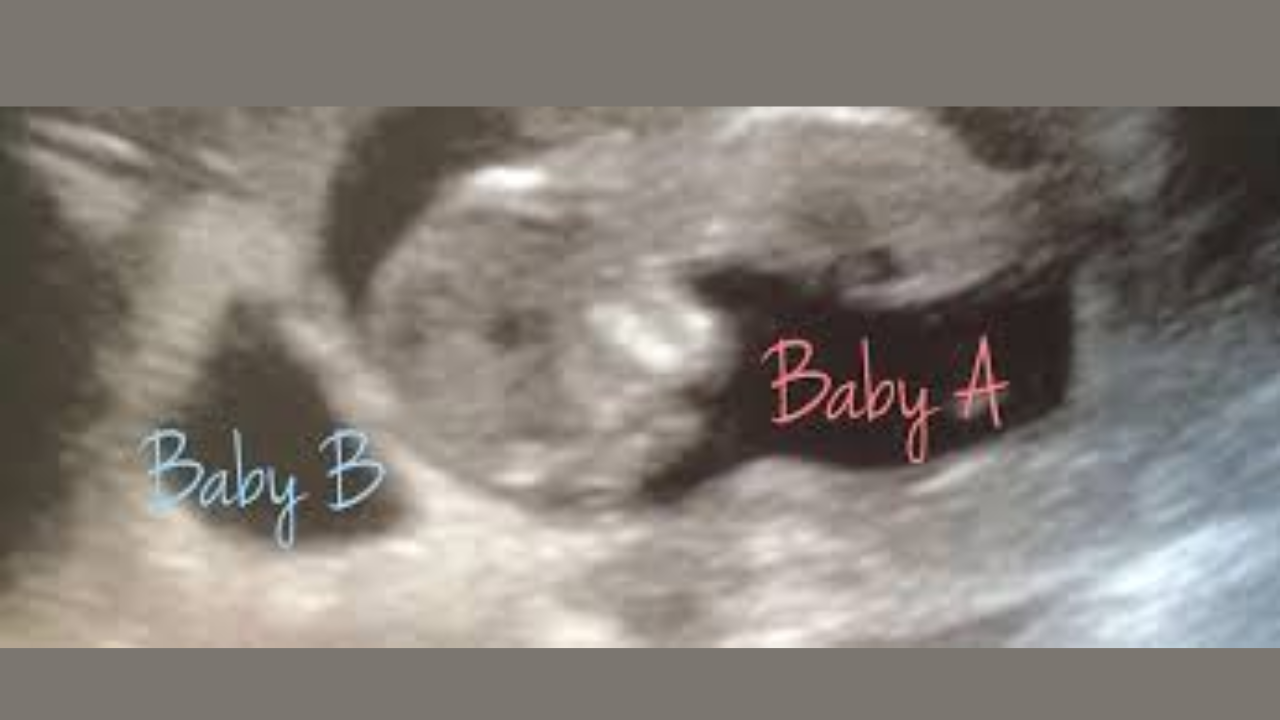
Love Island’s Olivia Bowen Pregnancy Update: Shares How She Grappled With News Of Vanishing Twin Syndrome

(Credit-Olivia Bowen/Instgaram)
Vanishing Twin Syndrome: The former Love Island contestant Olivia Bowen has recently shared a pregnancy update, following her post about her struggles to grapple with the reality that she will no longer be having twins, but just one healthy young baby.
Within a few weeks of her sharing her twin pregnancy with her fans, she followed up with another post where she broke the devastating news of finding only one baby in the womb.
In the caption she said, “The crazy sickness, the biggest surprise of our lives finding out we were having twins, imagining our lives with two new babies, then the complete heartache of dealing with vanishing twin syndrome & losing one of our babies”.
However, as devastating as the news may be, it is important to note that this is a more common occurrence than people realize. The National Institute of Health, US, statistics show that half of the pregnancies with three gestational sacs go through it and 36% of twin pregnancies also experience this. But what exactly is Vanishing Twin Syndrome?
Also Read: Walking Dead Actress Kelley Mack Dies At 33 After Battling Glioma
What Is Vanishing Twin Syndrome?
According to the American Pregnancy Association vanishing twin syndrome was first recognized in 1945. It's what happens when one of two or more babies in a pregnancy dies in the womb. The other twin, the placenta, or the mother's body then absorbs the dead baby's tissue. This makes it look like one of the babies has "vanished."
Thanks to early ultrasounds, doctors can now spot this more often. It's believed that vanishing twin syndrome happens in about 21-30% of pregnancies with more than one baby.
Causes and Signs of Vanishing Twin Syndrome
Most of the time, doctors don't know exactly why vanishing twin syndrome happens. However, they've found that the baby who is lost often had chromosomal problems, while the surviving twin is usually healthy. It seems these problems are there from the very beginning of the pregnancy. Another possible cause is that the umbilical cord didn't attach correctly. The signs of a possible vanishing twin syndrome usually happen early in the first trimester. They can include:
- Bleeding
- Cramping in the uterus
- Pain in the pelvis
- Research shows that this syndrome is more common in women over the age of 30.
How Does The Vanishing Twin Syndrome Affect The Mother And Baby?
If a twin is lost in the first three months of pregnancy, the surviving baby and the mother are usually fine. The living twin's chances of being healthy are very good.
If a twin is lost in the second or third trimester, there can be more risks for the surviving baby, including a higher chance of developing cerebral palsy. In these cases, the dead twin's body can get flattened by the pressure of the growing, healthy twin. At birth, doctors might find this flattened twin, which they call fetus compressus or fetus papyraceous.
How Do You Diagnoses Vanishing Twin Syndrome?
In the past, doctors could only figure out if a twin had died by looking at the placenta after the baby was born. Now, an early ultrasound can show twins in the first trimester. A later ultrasound might then show that one of the babies is no longer there. For example, a woman might see two heartbeats at 7 weeks but only one at her next visit.
If a twin is lost in the first trimester, no special medical care is usually needed for the mother or the surviving baby. However, if the death happens in the second or third trimester, the pregnancy may be treated as high-risk.
If you are pregnant and experience bleeding, cramping, or pelvic pain, you should see a doctor. An ultrasound will help them determine if a fetus is still viable before considering any procedures.
Leptospirosis: US health officials flag outbreak risk after floods in Hawaii
Credit: iStock
Health officials in the US have issued a risk of leptospirosis — a bacterial illness — in Hawaii, after the “Kona low” storm floods.
The Hawaii Department of Health (DOH) and the Hawaiian Humane Society (HHS) have urged residents and recovery workers to monitor for signs of leptospirosis that can affect both people and pets.
Leptospirosis is caused by bacteria Leptospira, which are carried in the urine of infected animals, including rats, mice, mongoose, livestock, and dogs.
The disease can spread via the urine of infected animals and can enter the human body through broken skin or through the mouth, nose, or eyes.
“Hawaii already has among the highest rates of leptospirosis in the country due to its warm, wet climate,” said HHS and DOH in a joint statement.
“Flooding events like the recent Kona low storm can dramatically increase exposure risk by spreading contaminated soil and water across a broad area,” it added.
The agency urged people to check out symptoms in pets, such as fever, decreased appetite, vomiting, diarrhea, and muscle soreness, and get medical help.
The risk of Leptospirosis outbreak is common after a storm, because the dead animals are likely to spread the bacteria. Further, flooding also forces rodents out of their burrows, leading them into homes, shelters, and debris piles where they shed bacteria more easily.
What Is Leptospirosis?
Leptospirosis is one of the most prevalent zoonotic diseases worldwide, often linked to poor sanitation and agricultural practices that involve contact with animals or contaminated water.
The bacteria can survive in contaminated water or soil for weeks to months, primarily spreading through the urine of infected animals.
Humans can contract the disease through direct contact with infected urine or through contaminated food, water, or soil.
There are two main forms of leptospirosis: icteric leptospira, which presents with jaundice, and anicteric leptospira, a milder variant without jaundice. Leptospirosis can also manifest as Weil's disease, a severe complication.
What Are Symptoms Of Leptospirosis in humans?
Symptoms typically develop 2 to 30 days after exposure and can range from mild to severe. Initial signs often include
- high-grade fever,
- red eyes (conjunctival suffusion),
- calf tenderness,
- headache,
- dry cough,
- nausea,
- diarrhea.
In more serious cases, the infection can lead to complications such as
- jaundice,
- kidney damage
- liver damage,
- meningitis,
- respiratory issues,
- hemorrhages.
Pulmonary hemorrhage — acute bleeding from the lungs — can also occur, posing life-threatening risks such as respiratory and renal failure. The illness may last from a few days to several weeks, and without treatment, recovery can take months, according to the US Centers for Disease Control and Prevention (CDC).
What Precautions Should Be Taken?
To reduce the risk of leptospirosis infection, individuals are advised to take several important precautions. According to the CDC, it is essential to avoid wading through dirty rainwater, especially during the rainy season, and to wear gumboots in such conditions.
Proper care of any injuries or cuts is crucial, and frequent handwashing, particularly after handling food or working in potentially contaminated environments, is highly recommended. Additionally, individuals should avoid contact with animals that appear sick or are known carriers of the bacteria.
Vaccination is also vital—pet owners should ensure their pets are vaccinated against leptospirosis, especially if they are frequently exposed to water or wildlife. Staying hydrated by drinking plenty of clean water and preventing water stagnation around the home can further minimize the risk of infection.
Cicada COVID Variant: Use Masks To Avoid Transmission, Say Experts

Credit: Canva
The emerging COVID variant, BA 3.2, nicknamed “Cicada,” has revived memories of the COVID-19 pandemic that disrupted the world and raised fresh concerns about the possibility of severe illness.
The variant has been given the nickname “Cicada” due to its reappearance after remaining dormant or undetected for a long period, much like cicadas that emerge after years underground.
With the variant already spread to 23 nations, as of February, experts are urging people to use masks to avoid transmission.
What Is The Cicada COVID Variant?
Cicada was first identified in a respiratory sample in South Africa in November 2024.
It is a descendant of the Omicron BA.3 lineage, and is genetically distinct from the previously circulating JN.1 lineages (including LP.8.1 and XFG).
BA.3.2 comprises two major branches, BA.3.2.1 and BA.3.2.2. BA.3.2.2 also has substitutions like: K356T, A575S, R681H, and R1162P.
The World Health Organization (WHO) has designated BA.3.2 as a Variant Under Monitoring (VUM). It means the variant may not be that dangerous yet, but it may have concerning mutations.
As per the US CDC’s latest Morbidity and Mortality Weekly Report, Cicada has “70 to 75 substitutions and deletions in the gene sequence of its spike protein”.
Time To Mask Up
Dr. Sai Balasubramanian, a doctor and healthcare strategy executive, writing in Forbes, stressed the need to follow COVID practices such as masks and hand hygiene.
"Healthcare professionals recommend taking general precautions, similar to those used to prevent most viral transmission: get vaccinations when appropriate, wear masks in crowded areas or indoors where there is a high risk of transmission," he said.
He also urged “avoid individuals who have known illness or infections, wash hands frequently, and continue to stay apprised of local community guidelines and the infection spread”.
Cicada variant “is different from the (Covid-19) viruses we have been dealing with for the last two years," Prof Ravi Gupta, of Cambridge University, who advised the UK government during the pandemic, was quoted as saying by The Mirror.
Will The Cicada Variant Cause Severe Illness?
The SARS-CoV-2 virus has the potential to turn deadly in people, especially among vulnerable populations such as those with a weak immune system.
The Cicada variant is particularly concerning as it provides no immune protection to people with previous infection or even vaccination.
Yet, the World Health Organization and health experts advise that existing COVID vaccinations can help prevent severe illness and hospitalization.
"It would appear that all the protections we have from our experience with the virus and with vaccines probably offer more limited—not zero—but more limited protection against this strain," Dr. William Schaffner, professor of infectious diseases at Vanderbilt University Medical Center, was quoted as saying by Time.
Cicada Variant: Any New Symptoms?
The symptoms of Cicada aren't different from those of previous COVID variants. These include:
- sore throat,
- fever or chills,
- headache,
- cough,
- body aches,
- runny nose
- nausea
- diarrhea.
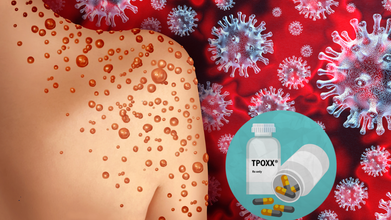
Tpoxx Should No Longer Be Used For Treating Mpox: European Drug Regulator
Credit: Canva/ US CDC
The European Medicines Agency (EMA) has advised patients and physicians not to use the antiviral drug Tecovirimat SIGA (marketed as Tpoxx) to treat mpox disease.
The advisory from the EMA’s Committee for Human Medicines (CHMP) is based on data from four studies carried out in different regions, which showed that compared with placebo treatment with Tpoxx did not
- heal lesions faster
- relieve pain
- help clear the virus from the body faster.
“The animal data demonstrated antiviral activity and a survival benefit when treatment was started early and a reduced efficacy if treatment was initiated later after exposure to the virus,” the EMA said in an official statement.
However, the clade 2 mpox outbreaks, which began in 2022, led the World Health Organization (WHO) to declare mpox a Public Health Emergency of International Concern (PHEIC), giving the drugmaker SIGA Technologies an opportunity to conduct efficacy studies in people.
A second PHEIC was declared in 2024 over clade 1 outbreaks.
Key recommendations by the EMA include:
- Restricting the use of Tpoxx over no benefit.
- Due to lack of safety concerns, patients who initiated treatment with Tpoxx can continue till course ends.
- Tpoxx can still be used as an antiviral to treat other orthopoxvirus infections, including smallpox and cowpox
- There is currently no other drug authorized in the EU for treating mpox infections.
Tpoxx For Mpox: What Does The US CDC Say
The Centers for Disease Control and Prevention also noted that the role of Tpoxx in treating mpox "is investigational".
The federal agency called for "additional clinical trials" to explore the role of Tpoxx in treating mpox infections in patients with severe immunocompromise, including advanced HIV.
"The findings from the clinical trials suggest that most patients with monkeypox who do not have severe disease or risk factors for severe disease (e.g., severe immunocompromise) will recover with supportive care and pain management," the CDC said.
Also read: Missouri Reports 2 Cases Of Deadly Clade I Mpox, US Tally Rises To 3 In 2026
What Is Mpox?
Mpox, earlier known as monkeypox, is a rare viral disease that belongs to the orthopoxvirus genus, the same family as the more well-known smallpox virus.
Though generally less severe, mpox can still lead to serious health complications, especially in immunocompromised individuals, children, and pregnant women.
The virus is transmitted from animals to humans and can spread from person to person through direct contact with infectious sores, scabs, body fluids, respiratory droplets, or contaminated materials.
Also read: Mpox Outbreak: First Case of Severe Strain Reported in New York City
The earliest signs of mpox start within 14 days of being infected. A person may not know they have mpox and can spread the disease.
The common symptoms include:
- fever,
- sweating,
- chills through the body.
- rashes, which start as a distant rash on the face and can continue throughout the body,
- swollen lymph nodes, migraine,
- muscle aches,
- fatigue,
- weakness
- back pain.
Mpox: Current Cases
In February, a total of 1,184 confirmed mpox cases and four deaths were reported from 46 countries, as per the WHO's latest outbreak update.
Of these cases, 58.6 percent were reported in Africa — mainly from Madagascar, the Democratic Republic of the Congo, Kenya, Burundi, and Liberia.
The WHO said all clades continue to circulate, and transmission of the virus continues mostly within sexual networks, followed by household transmission. All age-groups in some historically endemic areas are being affected.
“Unless mpox outbreaks are rapidly contained and human-to-human transmission is interrupted, there is a risk of sustained community transmission in all settings,” the WHO said.
- Follow Us :


© 2024 Bennett, Coleman & Company Limited