- Health Conditions A-Z
- Health & Wellness
- Nutrition
- Fitness
- Health News
- Ayurveda
- Videos
- Medicine A-Z
- Parenting
Walking Dead Actress Kelley Mack Dies At 33 After Battling Glioma

Credits: Instagram
Walking Dead famed actress Kelley Mack has passed away at the age of 33 on Saturday at her birthplace of Cincinnati. Her demise was confirmed by a social media post by her sister, who mentioned, "It is with indelible sadness that we are announcing the passing of our dear Kelley. Such a bright, fervent light has transitioned to the beyond, where we all eventually must go."
As per Deadline, the actress was battling with glioma of the central nervous system. She had also posted her health update battling the same, and shared how her radiation therapy had been going.
What Is Glioma?
As per the Johns Hopkins Medicine, glioma is a common type of tumor originating in the brain. About 33% of all brain tumors are gliomas, which originate n the glial cells that surround and support neurons in the brains, including astrocytes, oligodendrocytes and ependymal cells.
Gliomas are called intra-axial brain tumors because they grow within the substance of the brain and often mix with normal brain tissue.
Types Of Glioma
Astrocytomas: Astrocytomas are tumors that form in glial cells, specifically from connective tissue cells called astrocytes. They are the most common type of primary intra-axial brain tumor, making up almost half of all primary brain tumors. These tumors are most often found in the cerebrum, the large outer part of the brain, but they can also occur in the cerebellum, located at the base of the brain.
Astrocytomas can affect both children and adults. In children, low-grade astrocytomas, known as pilocytic astrocytomas, are typically found in the cerebellum. In adults, these tumors are more commonly located in the cerebrum. The most aggressive form of astrocytoma is glioblastoma multiforme, a high-grade tumor that is considered the most malignant of all brain tumors. Its symptoms are often similar to those seen with other types of gliomas.
Brain stem gliomas: Brain stem gliomas, also known as diffuse infiltrating brainstem gliomas or DIPGs, are rare and usually found in the brain stem. Due to their location and the way they grow by blending into normal brain tissue, they are generally not removable by surgery. These tumors most often affect school-age children and are responsible for the highest number of childhood deaths from primary brain tumors.
Ependymomas: Ependymomas develop from ependymal cells, which line the brain’s ventricles or the spinal cord. Although they are rare, making up just 2 to 3 percent of primary brain tumors, they account for 8 to 10 percent of brain tumors in children, especially those younger than 10. In children, these tumors are usually found near the cerebellum, where they may block the flow of cerebrospinal fluid and lead to increased pressure in the skull. Ependymomas can also spread to other areas of the brain or spinal cord through spinal fluid, a process known as drop metastasis.
Mixed gliomas: Mixed gliomas, also called oligoastrocytomas, contain more than one type of glial cell. There is some debate over whether these tumors should be classified as a separate type, and genetic testing of tumor tissue is often used to clarify the diagnosis. These tumors usually occur in the cerebrum and are most common in adult men.
Oligodendrogliomas : Oligodendrogliomas form from oligodendrocytes, the supportive cells in the brain, and are usually located in the cerebrum. They make up about 2 to 4 percent of primary brain tumors and are more common in men, especially those in young to middle adulthood. Seizures are a frequent symptom, affecting up to 80 percent of patients, along with headaches, weakness, or speech problems. Oligodendrogliomas tend to have a better prognosis than many other gliomas.
Optic pathway gliomas: Optic pathway gliomas are low-grade tumors found in the optic nerve or optic chiasm. These tumors often affect people with neurofibromatosis and can lead to vision loss and hormone-related problems, especially since they tend to grow at the base of the brain, near areas responsible for hormone control. When these tumors impact hormone function, they may be referred to as hypothalamic gliomas.
What Are The Symptoms of Glioma?
- Headaches
- Seizures
- Personality changes
- Weakness in the arms, face or legs
- Numbness
- Problems with speech
- Nausea and vomiting
- Vision loss
- Dizziness
Cicada COVID Variant: Use Masks To Avoid Transmission, Say Experts

Credit: Canva
The emerging COVID variant, BA 3.2, nicknamed “Cicada,” has revived memories of the COVID-19 pandemic that disrupted the world and raised fresh concerns about the possibility of severe illness.
The variant has been given the nickname “Cicada” due to its reappearance after remaining dormant or undetected for a long period, much like cicadas that emerge after years underground.
With the variant already spread to 23 nations, as of February, experts are urging people to use masks to avoid transmission.
What Is The Cicada COVID Variant?
Cicada was first identified in a respiratory sample in South Africa in November 2024.
It is a descendant of the Omicron BA.3 lineage, and is genetically distinct from the previously circulating JN.1 lineages (including LP.8.1 and XFG).
BA.3.2 comprises two major branches, BA.3.2.1 and BA.3.2.2. BA.3.2.2 also has substitutions like: K356T, A575S, R681H, and R1162P.
The World Health Organization (WHO) has designated BA.3.2 as a Variant Under Monitoring (VUM). It means the variant may not be that dangerous yet, but it may have concerning mutations.
As per the US CDC’s latest Morbidity and Mortality Weekly Report, Cicada has “70 to 75 substitutions and deletions in the gene sequence of its spike protein”.
Time To Mask Up
Dr. Sai Balasubramanian, a doctor and healthcare strategy executive, writing in Forbes, stressed the need to follow COVID practices such as masks and hand hygiene.
"Healthcare professionals recommend taking general precautions, similar to those used to prevent most viral transmission: get vaccinations when appropriate, wear masks in crowded areas or indoors where there is a high risk of transmission," he said.
He also urged “avoid individuals who have known illness or infections, wash hands frequently, and continue to stay apprised of local community guidelines and the infection spread”.
Cicada variant “is different from the (Covid-19) viruses we have been dealing with for the last two years," Prof Ravi Gupta, of Cambridge University, who advised the UK government during the pandemic, was quoted as saying by The Mirror.
Will The Cicada Variant Cause Severe Illness?
The SARS-CoV-2 virus has the potential to turn deadly in people, especially among vulnerable populations such as those with a weak immune system.
The Cicada variant is particularly concerning as it provides no immune protection to people with previous infection or even vaccination.
Yet, the World Health Organization and health experts advise that existing COVID vaccinations can help prevent severe illness and hospitalization.
"It would appear that all the protections we have from our experience with the virus and with vaccines probably offer more limited—not zero—but more limited protection against this strain," Dr. William Schaffner, professor of infectious diseases at Vanderbilt University Medical Center, was quoted as saying by Time.
Cicada Variant: Any New Symptoms?
The symptoms of Cicada aren't different from those of previous COVID variants. These include:
- sore throat,
- fever or chills,
- headache,
- cough,
- body aches,
- runny nose
- nausea
- diarrhea.
Tpoxx Should No Longer Be Used For Treating Mpox: European Drug Regulator
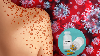
Credit: Canva/ US CDC
The European Medicines Agency (EMA) has advised patients and physicians not to use the antiviral drug Tecovirimat SIGA (marketed as Tpoxx) to treat mpox disease.
The advisory from the EMA’s Committee for Human Medicines (CHMP) is based on data from four studies carried out in different regions, which showed that compared with placebo treatment with Tpoxx did not
- heal lesions faster
- relieve pain
- help clear the virus from the body faster.
“The animal data demonstrated antiviral activity and a survival benefit when treatment was started early and a reduced efficacy if treatment was initiated later after exposure to the virus,” the EMA said in an official statement.
However, the clade 2 mpox outbreaks, which began in 2022, led the World Health Organization (WHO) to declare mpox a Public Health Emergency of International Concern (PHEIC), giving the drugmaker SIGA Technologies an opportunity to conduct efficacy studies in people.
A second PHEIC was declared in 2024 over clade 1 outbreaks.
Key recommendations by the EMA include:
- Restricting the use of Tpoxx over no benefit.
- Due to lack of safety concerns, patients who initiated treatment with Tpoxx can continue till course ends.
- Tpoxx can still be used as an antiviral to treat other orthopoxvirus infections, including smallpox and cowpox
- There is currently no other drug authorized in the EU for treating mpox infections.
Tpoxx For Mpox: What Does The US CDC Say
The Centers for Disease Control and Prevention also noted that the role of Tpoxx in treating mpox "is investigational".
The federal agency called for "additional clinical trials" to explore the role of Tpoxx in treating mpox infections in patients with severe immunocompromise, including advanced HIV.
"The findings from the clinical trials suggest that most patients with monkeypox who do not have severe disease or risk factors for severe disease (e.g., severe immunocompromise) will recover with supportive care and pain management," the CDC said.
Also read: Missouri Reports 2 Cases Of Deadly Clade I Mpox, US Tally Rises To 3 In 2026
What Is Mpox?
Mpox, earlier known as monkeypox, is a rare viral disease that belongs to the orthopoxvirus genus, the same family as the more well-known smallpox virus.
Though generally less severe, mpox can still lead to serious health complications, especially in immunocompromised individuals, children, and pregnant women.
The virus is transmitted from animals to humans and can spread from person to person through direct contact with infectious sores, scabs, body fluids, respiratory droplets, or contaminated materials.
Also read: Mpox Outbreak: First Case of Severe Strain Reported in New York City
The earliest signs of mpox start within 14 days of being infected. A person may not know they have mpox and can spread the disease.
The common symptoms include:
- fever,
- sweating,
- chills through the body.
- rashes, which start as a distant rash on the face and can continue throughout the body,
- swollen lymph nodes, migraine,
- muscle aches,
- fatigue,
- weakness
- back pain.
Mpox: Current Cases
In February, a total of 1,184 confirmed mpox cases and four deaths were reported from 46 countries, as per the WHO's latest outbreak update.
Of these cases, 58.6 percent were reported in Africa — mainly from Madagascar, the Democratic Republic of the Congo, Kenya, Burundi, and Liberia.
The WHO said all clades continue to circulate, and transmission of the virus continues mostly within sexual networks, followed by household transmission. All age-groups in some historically endemic areas are being affected.
“Unless mpox outbreaks are rapidly contained and human-to-human transmission is interrupted, there is a risk of sustained community transmission in all settings,” the WHO said.
Study Shows Ebola Virus May Persist In Breast Milk For Over 3 Months After Recovery
Credit: Canva
Even after recovery, the deadly Ebola virus (EBOV) can persist for a longer duration — more than three months — in breastmilk, according to a case report.
Ebola virus disease (EVD) is a severe viral illness that has a 25 percent – 90 percent fatality rate.
Ebola in pregnancy raises significant complications ranging from spontaneous abortion to maternal and neonatal death.
In a case report published in the New England Journal of Medicine, a team of researchers from the Republic of Congo and Senegal shared the case history of a 23-year-old woman in whom Ebola was still detectable in breast milk at 14 weeks.
The case reported the rare occurrence of a pregnant woman who survived EVD with no complications, neither to the mother nor the baby. However, the deadly virus was still present in the mothers' breast milk, which cited the potential risk of post-illness transmission to infants.
What Is Ebola Virus Disease?
As per the World Health Organization (WHO), EVD is a rare but severe illness in humans and is often fatal.
People can get infected with the virus if they touch an infected animal when preparing food, or touch body fluids of an infected person such as saliva, urine, feces or semen, or things that have body fluids of an infected person like clothes or sheets.
Ebola enters the body through cuts in the skin or when one is touching their eyes, nose or mouth. Early symptoms include fever, fatigue and headache.
What Was The Case
The woman from the Democratic Republic of the Congo (DRC) contracted EBOV during pregnancy in 2019.
Soon after, she was administered monoclonal antibody therapy and was discharged after three negative reverse-transcriptase–polymerase-chain-reaction blood tests for EBOV.
The woman delivered a healthy baby at 42 weeks of gestation. No evidence of EBOV infection was found in maternal blood, amniotic fluid, vaginal secretions, or the newborn.
Ebola Detectable In Breast Milk At 14 Weeks
Yet surprisingly, the EBOV virus persisted in the placenta and breast milk.
Tests revealed that while the mother’s blood remained negative, viral RNA was still detectable in breast milk at 14 weeks after illness onset. To protect the newborn from transmission, clinicians used the drug bromocriptine to suppress lactation.
As per the World Health Organization (WHO) guidelines, the mother was also isolated from the baby and not breastfed. A prophylactic (preventive) monoclonal antibody was also given to the newborn. During follow-up, the infant exhibited no signs of infection.
Ebola Virus: WHO Guidelines
During the 2018–2020 EVD outbreak in the northeast of DRC, 3,481 confirmed cases were reported. Of these, nearly 60 percent occurred in females, and about 45 percent occurred in children below 18 years of age.
Current WHO guidance recommends that Ebola survivors avoid breastfeeding until viral clearance is confirmed.
The global health body advises women with suspected or confirmed Ebola to immediately stop breastfeeding and be prioritized for diagnostic testing.
Children exposed to Ebola through breast milk
- Must be placed under care,
- Closely monitored for symptoms over 21 days
- Fed with an appropriate breast milk substitute.
- Restarted on breastfeeding after two consecutive negative tests of breast milk.
- Follow Us :


© 2024 Bennett, Coleman & Company Limited