- Health Conditions A-Z
- Health & Wellness
- Nutrition
- Fitness
- Health News
- Ayurveda
- Videos
- Medicine A-Z
- Parenting
Why Placenta Banking Is Being Called the Ultimate Health Insurance for Families

Credits: Canva
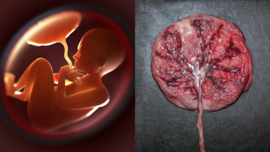
If you thought the only souvenirs from childbirth were baby pictures and tiny socks, times have changed. Turns out, the real treasure might be something most parents never even glance at before it is thrown away: the placenta and umbilical cord. Doctors are now calling placenta banking “biological insurance”, and the idea is picking up pace.
Your Baby’s Placenta Is More Than Just Leftovers
For centuries, the placenta has been treated as medical waste. But according to Dr. D.B. Usha Rajinikanthan, Senior Consultant in Gynaecology and IVF at SIMS Hospital, Chennai, this organ is brimming with stem cells that could be life-saving later on.
“Placenta and cord blood contain stem cells that can repair or replace damaged tissue. Collecting them at birth is safe and painless, but once discarded, that opportunity is lost forever,” she says.
These tiny cells are essentially the body’s master builders, with the potential to transform into different blood and immune cells. Which means what is usually thrown in a bin could actually hold a family’s medical safety net.
Why Stem Cells Are a Big Deal
Stem cells from the placenta are not just versatile; they are generous. Dr. Rajinikanthan explains that they have already been used to treat more than 80 diseases worldwide, including leukaemia, certain immune deficiencies and metabolic disorders. “Research is expanding into conditions like heart repair, brain injury and even diabetes,” she adds.
Placental stem cells are “younger” and more flexible, making them easier to match with siblings and relatives. In simple terms, the baby, siblings, parents and even grandparents may stand to benefit. It is not just your child’s resource; it is potentially a family heirloom.
Placenta Preservation: A Health Insurance
If we insure our cars and houses against accidents, why not our health? Placenta banking works on that philosophy. “It is a one-time investment in future health security. Families may never need it, but having stored stem cells gives enormous peace of mind,” says Dr. Rajinikanthan. She emphasises, though, that choosing an accredited stem cell bank that follows quality standards is essential.
How Does Amniotic Membrane Help?
Beyond the cord blood, there is another underrated star, the amniotic membrane. Dr. A. Jaishree Gajaraj, Head of Obstetrics and Gynaecology at MGM Healthcare, Chennai, explains that the amnion has been saving lives for over a century. “The first use dates back to 1910 when it was applied as a skin graft to promote healing. Today, it is used in ophthalmology for dry eyes, as well as for burns and diabetic ulcers,” she says.
In other words, this part of the placenta is not just a wrapper for your baby; it is a medical toolkit waiting to be tapped.
The Science Behind the Promise
Stem cell science has moved leaps and bounds in recent decades. According to Dr. Gajaraj, the umbilical cord blood and tissue have already been used successfully in bone marrow transplants for children with leukaemia and other bone marrow disorders. But the real buzz is around their future potential.
“These pluripotent cells are being researched for regenerating organs like the pancreas, liver, lungs and even the spinal cord. While still experimental, the promise is extraordinary,” she explains.
She adds that mesenchymal stem cells (MSCs), particularly those derived from cord tissue, are showing the greatest promise in regenerative therapies. “Foetal MSCs from cord tissue expand better, are less likely to trigger immune rejection, and have higher therapeutic potential than their maternal counterparts,” says Dr. Gajaraj. Simply put, storing placenta and cord tissue maximises the number and types of cells available for future therapies.
But What About Delayed Cord Clamping?
Some parents worry that opting for placenta banking might compromise delayed cord clamping, the practice of waiting a few minutes before cutting the cord to allow extra blood flow to the baby. Dr. Gajaraj reassures that this is not the case. “Delayed clamping does not reduce the yield of mesenchymal stem cells. Parents can safely choose both practices,” she says.
A Gift From Your Newborn to the Whole Family
Placenta banking is not a crystal ball or a cure-all. It does not guarantee immunity against every illness. But as both doctors point out, it offers a shot at future treatments that could transform outcomes in life-threatening conditions
Can The HPV Vaccine Impact Your Chances Of Conceiving? Expert Answers

Credits: Canva
Concerns around fertility and vaccines often surface when people plan a family, and the HPV vaccine is no exception. Many women and men worry that getting vaccinated today could affect their ability to conceive later in life. Medical experts, however, say this fear is misplaced. According to fertility specialists, there is no evidence linking the HPV vaccine to reduced fertility. In fact, the vaccine may play a quiet but important role in protecting reproductive health over the long term.
Does The HPV Vaccine Affect Fertility?
The short and clear answer is no. The HPV vaccine does not negatively affect fertility in women or men. Dr. Madhu Patil, Consultant and Fertility Specialist at Motherhood Fertility and IVF, Sarjapur, Bangalore, explains that there is no scientific proof showing the vaccine causes fertility problems of any kind.
She notes that concerns often arise from misinformation rather than medical data. Extensive research and global vaccination programmes have consistently shown that people who receive the HPV vaccine do not experience reduced chances of conceiving in the future.
How HPV Infection Can Threaten Future Fertility
While the vaccine itself does not harm fertility, an untreated HPV infection can. HPV is the leading cause of nearly all cervical cancer cases. As per Dr Patil, “treatment for cervical cancer often involves procedures such as cone biopsy or LEEP, which can weaken the cervix. In more advanced cases, radiation or chemotherapy may be required.”
These treatments can reduce a woman’s ability to conceive and, in some cases, make it difficult to carry a pregnancy to full term. By preventing HPV-related cancers in the first place, the vaccine helps preserve the reproductive system and lowers the risk of fertility-compromising treatments later in life.
Why The HPV Vaccine Supports Reproductive Health
Dr. Patil points out that the HPV vaccine should be viewed as a protective measure rather than a risk. By stopping high-risk HPV strains from causing cancer or precancerous changes, the vaccine helps maintain cervical health. A healthy cervix and reproductive system are key factors in natural conception and safe pregnancies.
In this way, the vaccine indirectly supports fertility by reducing the likelihood of medical interventions that could interfere with reproductive function.
When Should the HPV Vaccine Be Taken?
Health experts recommend starting HPV vaccination at ages 11 or 12. At this stage, the immune response is strongest, and the vaccine offers protection well before any potential exposure to the virus. Dr. Patil strongly encourages parents to consult a gynaecologist and consider timely vaccination for their children.
That said, adults who missed vaccination earlier can still benefit. Many women and men receive the vaccine later in life after discussing it with their doctor.
Why Men Should Also Get The HPV Vaccine
The HPV vaccine is not only for women. Dr. Patil stresses that men should also be vaccinated, as HPV can cause cancers and genital warts that affect sexual health. Vaccination in men also reduces transmission to partners, adding another layer of protection for couples planning a family.
By limiting the spread of HPV, vaccination helps safeguard the reproductive and sexual health of both partners.
There is no evidence that the HPV vaccine reduces fertility. On the contrary, it helps prevent cancers and medical treatments that can threaten the ability to conceive or carry a pregnancy. Experts advise speaking with a gynaecologist, understanding the benefits, and making an informed decision based on medical facts rather than fear.
Toddler Left Partially Blind After ‘Ear Infection’ Diagnosis—What Did Doctors Miss?

Credits: Canva
A three-year-old girl was left partially blind after what first seemed like a routine ear infection was later diagnosed as a life-threatening brain tumour. As per Express UK, Chloe Kefford was rushed to A&E when she started experiencing car sickness and balance problems. Doctors initially diagnosed her with an ear infection and sent her home with antihistamines. But as Chloe’s condition worsened, her parents insisted on further testing, which revealed a tumour affecting her optic nerve.
Ear Infection Symptoms Masked A Life-Threatening Diagnosis
Chloe, from Formby, Merseyside, underwent open brain surgery and faced three-and-a-half years of treatment, including proton beam therapy last year, after experiencing two relapses. Proton beam therapy uses high-energy protons to precisely target the tumour, limiting damage to surrounding healthy tissue.
Open Brain Surgery And Years Of Intensive Treatment Followed
Now nine years old, Chloe has been honoured with a special award from Cancer Research UK for her bravery throughout her treatment. She received her initial care at St George’s Hospital in London and The Royal Marsden, before being transferred to Alder Hey in Liverpool.
Proton Beam Therapy Used After Cancer Relapsed Twice
Chloe’s mother, Nikki, 38, recalled that the family had been planning a move from Surrey to Merseyside before Chloe fell ill. As per Express UK, she said: “The house was already sold and we were planning our new life by the beach when Chloe became ill. Then we ended up moving and having to isolate for months. She relapsed not long after we moved and had more chemotherapy, then she rang the bell in April last year, but unfortunately, she relapsed again in July. So, we were supposed to be going on holiday to Disneyland in Paris and instead we went to Manchester for six weeks for Chloe to have proton beam therapy.”
Nikki added: “She is partially sighted now and has no peripheral vision; one eye is particularly badly affected. The main aim now is to preserve what eyesight she has left. We’re hopeful that the recent targeted treatment has got the cancer once and for all. She’s on steroids at the moment and is being monitored with three-monthly scans. She’s still in recovery and struggles with fatigue from the treatment, but we hope she’ll have a bit more energy soon. She’s our little ray of sunshine.”
Each year, around 400 children and young people in the North West are diagnosed with cancer. Advances in treatment and research are helping make therapies more effective and less harmful. Alder Hey Children’s Hospital in Liverpool is one of several centres across the UK taking part in pioneering clinical trials offering innovative new treatments.
In 2018, Cancer Research UK launched the Children’s Brain Tumour Centre of Excellence, supported by TK Maxx. The virtual centre brings together international experts in children’s brain tumour research to transform how treatments are developed. Every child nominated for a Star Award receives this recognition, which is endorsed by celebrities including JoJo Siwa and Pixie Lott.
Cancer Research UK spokesperson Jemma Humphreys said: “After everything Chloe’s been through, it’s been an absolute privilege to celebrate her incredible courage with a Star Award.”
Norovirus Spreads Rapidly In UK With Doctors Flagging New Symptoms

Credits: Canva
People experiencing certain symptoms are being urged to stay at home as a highly contagious virus spreads quickly across England. Fresh figures from the UK Health Security Agency show a 47% rise in cases during the first two weeks of 2026. This sudden jump has led the agency to remind the public about basic hygiene steps that play a key role in limiting the spread. Data suggests that norovirus is affecting people aged 65 and above the most, and although overall activity remains within normal seasonal levels, there has been a noticeable increase in outbreaks in hospital settings.
The latest UKHSA surveillance update also points to falling levels of flu, COVID-19, and RSV in the opening week of the year. While all winter virus levels are currently where they would be expected for this time of year, people are being encouraged to continue following simple precautions to help keep infections on a downward path.
What Is Norovirus?
Norovirus is an extremely infectious virus that irritates the stomach and intestines, causing gastroenteritis. It often leads to sudden vomiting, diarrhoea, nausea, and stomach cramps, and in some cases may be accompanied by fever or body aches. Although it is sometimes referred to as the “stomach flu,” it has no link to influenza. The virus spreads easily through contaminated food or water, shared surfaces, or close contact with someone who is infected. According to the Cleveland Clinic, most otherwise healthy individuals recover within a few days with rest and fluids, but preventing dehydration and avoiding passing the virus on to others is essential.Norovirus Symptoms
Common symptoms of norovirus include:
- Nausea.
- Vomiting.
- Diarrhoea.
- Stomach pain.
You may also experience:
- Headache.
- Fever.
- Body aches.
Symptoms usually develop between 12 and 48 hours after exposure and typically last for one to three days.
Doctors Report New Symptoms Of Norovirus
Both flu and norovirus can behave unpredictably, with case numbers rising and falling throughout the season. This makes simple preventive steps especially important. For illnesses affecting the stomach or respiratory system, such as norovirus, regular handwashing remains one of the most effective measures.
Health experts stress that alcohol-based hand sanitisers do not work against norovirus. Washing hands thoroughly with soap and warm water, along with cleaning surfaces using bleach-based products, is far more effective in reducing the spread. Good ventilation indoors can also help limit the transmission of respiratory viruses like flu. Anyone who develops symptoms is advised to stay at home whenever possible.
If going out cannot be avoided, wearing a face covering may help, particularly when around people who are more vulnerable.
Amy Douglas, Lead Epidemiologist at the UKHSA, said, according to the Mirror: “We have seen a clear rise in norovirus cases in recent weeks, particularly among people aged 65 and over, alongside an increase in hospital outbreaks. Although levels are still within what we would normally expect, there are simple actions people can take to stop norovirus spreading further.
“Washing hands with soap and warm water and cleaning surfaces with bleach-based products are key steps. Alcohol gels do not kill norovirus, so they should not be relied on alone.
“If you have diarrhoea and vomiting, do not return to work, school, or nursery until 48 hours after symptoms have stopped, and avoid preparing food for others during this time. If you are unwell, please stay away from hospitals and care homes to protect those most at risk from infection.”
© 2024 Bennett, Coleman & Company Limited

