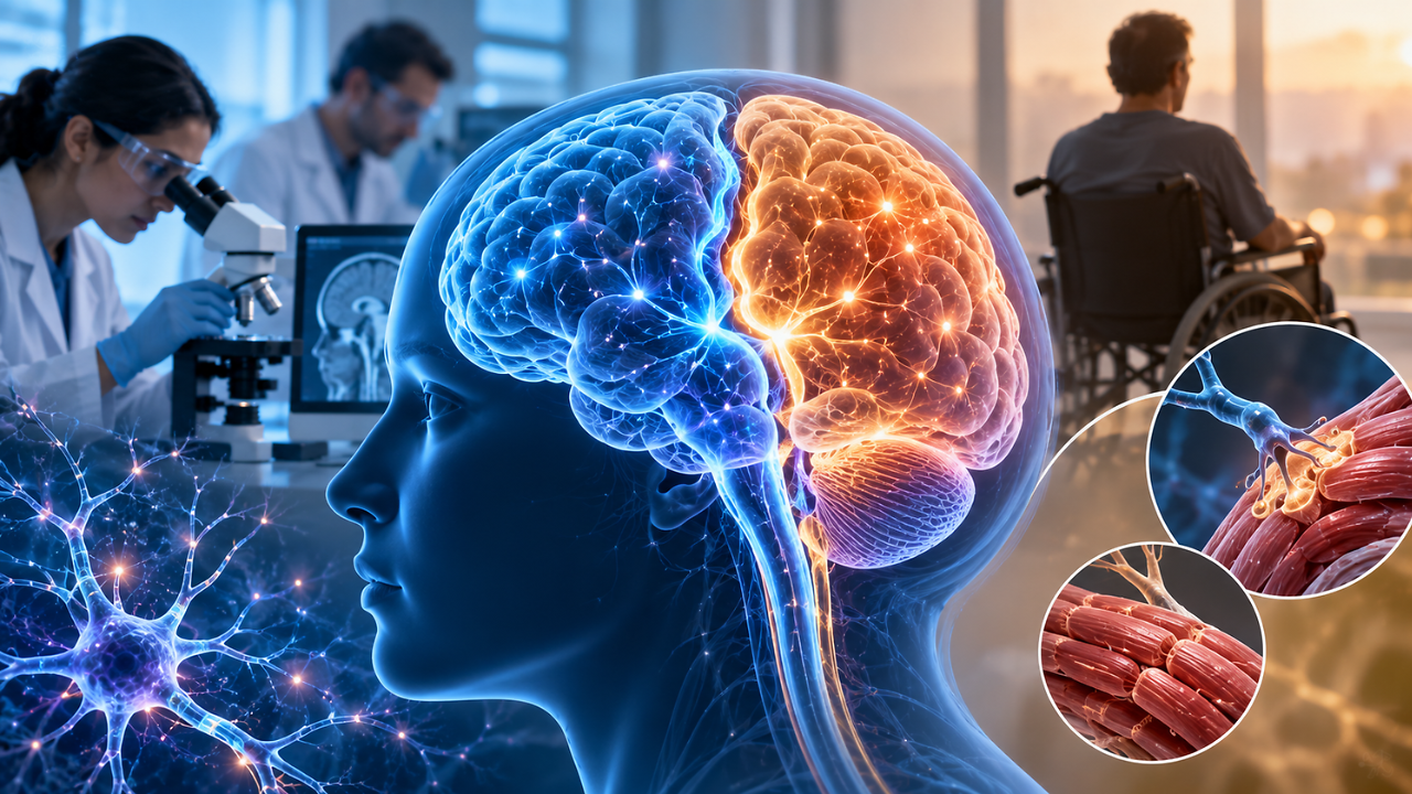
The human brain has always been considered to be a single organ. But according to Stanford Medicine researchers, the brain's growth history indicates that it originates from two different populations of early cells that follow separate developmental paths.The findings, published in Nature Neuroscience, suggest that the front and back parts of the brain develop from different types of progenitor cells, questioning the long-believed idea that the entire brain originates from one common cell.An important point to note is that this does not mean that humans have two completely separate brains. The two developmental systems ultimately fuse together and function as one brain.Mechanism Of Brain Forming From Two Cell GroupsDuring early development, cells called progenitor cells enable the production of specialised brain cells, including neurons. Researchers found that the cells that form the forebrain and midbrain follow a different developmental pathway from those that form the hindbrain, which includes the brainstem.The forebrain is involved in neurological functions like language, consciousness and higher-level thinking. The hindbrain contains structures that control essential functions including breathing, heartbeat, sleep and swallowing.Kyle Loh, associate professor of developmental biology at Stanford Medicine and senior author, said, “We’ve shown for the first time that the front of the brain arises from a totally different progenitor cell than the back of the brain.”He added, “Our discovery means that we can now grow neurons from the back of the brain, the hindbrain, in a petri dish and study their functions.”Also read: Is Controlling BP Enough After a Brain Bleed? Doctors Explain How to Prevent Another StrokeA Significant DiscoveryScientists have been able to grow several types of human brain cells in the laboratory, but creating hindbrain neurons has been challenging.The new findings give some clarity about why some previous approaches were unsuccessful. Researchers were attempting to transform forebrain or midbrain progenitor cells into hindbrain cells, but the Stanford team found that these cells are fundamentally different from the beginning of development.Jokhai, one of the researchers involved in the study, explained, “Previous attempts to make hindbrain neurons likely tried to coax forebrain and midbrain progenitors into hindbrain cells, which our study shows is not possible.”He added, “In stem cell biology, people are always fixated with creating the end cell type, like the neuron. But it’s important to begin at the earliest stages of embryonic development.”Also read: Brain Health After 40: How Diabetes, Menopause And Early Detection Affect Dementia Risk | World Alzheimer’s DayStudy's Implications On Brain Disease Research The discovery could have various practical implications for neurological research. The hindbrain and brainstem contain neurons involved in breathing, swallowing and movement, meaning researchers need reliable human cells to study diseases that damage these systems.Stanford scientists say the new findings allowed them to understand and generate functional human hindbrain neurons in the laboratory. This could provide new models for studying conditions like amyotrophic lateral sclerosis (ALS) and spinal muscular atrophy (SMA).Instead of studying these diseases only in animals or using cells that do not accurately represent the affected brain region, researchers may now be able to grow relevant human neurons and investigate how they develop, malfunction and respond to different treatments.The finding could therefore change not just how scientists understand the human brain's anatomy, but how they understand its origins and how they recreate different brain regions in the laboratory.