- Health Conditions A-Z
- Health & Wellness
- Nutrition
- Fitness
- Health News
- Ayurveda
- Videos
- Medicine A-Z
- Parenting
World Kidney Day 2025: Theme, History, and Significance

Credits: Canva
Every year on the second Thursday of March, we observe World Kidney Day to raise awareness about kidney health and its essential role in our overall well-being. This year, the day falls on March 13. Such health days are important as it highlights the need for paying attention to each part and organ of our body, which is essential to keep us going.
The kidneys are two bean-shaped organs which act as body's most essential and primary filtration system. They remove waste products and excess fluid from the blood.
The day is important to highlight because more often than you think, kidney diseases go unnoticed. Max Healthcare, a hospital chain in India states that 8 to 10% of adults are affected with some form of kidney damage. However, not many know about it, until severe complications arise. Each year, millions die prematurely from such conditions, this is why early detection is important as it can make a significant difference.
Theme For World Kidney Day
This is what has reflected in this year's theme for World Kidney Day: "Are Your Kidneys OK? Detect Early, Protect Kidney Health". This year's theme aims to target the importance of regular checkups for early detection and intervention at the right time to prevent any kidney disease.
As per the National Institutes of Health (NIH), the kidneys are important to maintain a healthy balance of water, salts, and minerals. These are extremely essential for regulating blood pressure, producing red blood cells, and ensuring overall body equilibrium.
What Happens When Your Kidney Fails?
When kidneys fail to perform its primary function, which is to filter out the waste and produce erythropoietin, which is a hormone that stimulates red blood cell production, it can lead to health complications. Your body gets filled with extra water and waste products, this condition is called uremia. As a result, your hands and feet may also swell. At the time of complete and irreversible kidney failure, your kidneys stop working completely. This condition is called End-stage renal disease or ESRD.
History of World Kidney Day
This day was for the first time observed in 2006 as a joint initiative by the Internal Society of Nephrology (ISN) and the International Federation of Kidney Foundations (IFKF). The aim back then too was to raise awareness about kidney health and to promote preventive measures to reduce the burden of kidney diseases worldwide. Since then, it has been observed on every second Thursday of March, every year.
Significance of World Kidney Day
The significance of World Kidney Day lies in its role in addressing the growing prevalence of kidney diseases. Many of the cases go undiagnosed. Other than that there are many cases among children where parents do not know whether it is a kidney disease or an unrelated infection. Thus this day also highlights the importance of paying attention to your child's health for any pediatric kidney issues. As this disease is often associated with adults only, but not always is this the case.
ALSO READ: Why Do Cases Of Pediatric Kidney Issues Go Unnoticed?
Becky Quick Speaks About Her Daughter’s Genetic Disorder —What The Diagnosis Is About

Credits: AP
Becky Quick recently spoke about her 9-year-old daughter Kaylie’s private struggle with a rare genetic disorder. In an interview with People, published Thursday, January 8, the 53-year-old co-anchor of CNBC’s Squawk Box revealed that Kaylie has SYNGAP1. According to the Child Neurology Foundation, SYNGAP1 is a genetic condition that can cause seizures and developmental challenges. Quick recalled that she first noticed something was off “probably around 8 months,” when Kaylie wasn’t reaching her developmental milestones.
“Sometimes her eyes would cross. You just know as a mom that something isn’t right,” Quick told People, adding that after observing her daughter and reviewing developmental studies, it became clear that Kaylie wasn’t meeting expected benchmarks. “We immediately began working with therapists. They supported Kaylie in rolling over, achieving fluid movements, eventually walking, and so many other skills. But we also noticed additional concerns developing.”
Becky Quick Daughter Battles SYNGAP1
For nearly a decade, Quick has kept her daughter Kaylie’s SYNGAP1 diagnosis private. The condition, as defined by the Children’s Hospital of Philadelphia, is a rare genetic disorder that can lead to a variety of neurodevelopmental challenges. It currently has no cure and often requires round-the-clock care.
The journalist took Kaylie to her pediatrician, who recommended a study with a developmental specialist. The results confirmed Quick’s fears: her daughter was not reaching key milestones.
“We started working with therapists immediately,” Quick said. “[They] helped Kaylie develop rolling, fluid movement, eventually walking, and many other skills. But we also observed additional issues.”
A neurologist then performed an EEG study, which revealed that Kaylie was experiencing subclinical seizures — subtle seizures that aren’t visibly noticeable. A subsequent genetic test confirmed that Kaylie had SYNGAP1.
What Is SYNGAP1?
SYNGAP1 (SRD) refers both to a gene and the rare neurological disorder it causes. The condition is marked by intellectual disability, epilepsy, and autism spectrum traits, arising from a mutation that reduces the SynGAP protein, which is essential for proper synaptic function in the brain, according to MedlinePlus. Without adequate SYNGAP1 protein, synapses become overly excitable, impairing communication between neurons and leading to the neurological difficulties observed in SYNGAP1 patients, as per National Institute of Health.
Becky Quick Daughter: What Are The Symptoms Of SRD?
SYNGAP1 is considered a spectrum disorder, meaning patients experience symptoms differently and with varying severity. The exact factors influencing symptom expression or intensity remain unclear. The most commonly observed signs include, as per NIH:
- Intellectual Disability (mild to severe)
- Hypotonia (low muscle tone)
- Global Developmental Delay
- Epilepsy (including subtle eyelid flutters, brief jerks, staring spells, and drop seizures)
- Sensory Processing Disorder
- Delays in Gross and Fine Motor Skills
- Dyspraxia (coordination difficulties)
- Speech Delay/Apraxia (mild to severe)
- Autism Spectrum Disorder
- Sleep and Behavior Disorders
- Visual Abnormalities
- Not every patient will exhibit all of these symptoms.
Becky Quick Daughter: What Causes SRD?
SYNGAP1-related disorders arise from a variant on the SYNGAP1 gene (6p.21.32). Each human cell contains 23 pairs of chromosomes, totaling 46, with thousands of genes on each chromosome. Most genes come in pairs, one inherited from each parent, and their primary role is to produce proteins that regulate tissues and organs.
When a gene experiences a variant, sometimes called a mutation or alteration — it can stop functioning properly. Variants occur naturally, similar to a typo when DNA is copied from cell to cell, or they may result from environmental factors.
A de novo variant is a genetic change that appears for the first time in a family, occurring early in reproductive development. Most SYNGAP1 patients carry de novo variants. For families with a genetic report, understanding the variant can help clarify the diagnosis and guide next steps.
India’s Antacid Habit Is Putting Hearts, Kidneys, Guts, and Bones at Risk

Credits: iStock
Indians are popping antacids like candies, and they are putting their hearts, kidneys and gut health at risk. Doctors from time and again have cautioned patients to not take such pills without prescriptions.
Antacids are prescribed proton pump inhibitors or PPIs, used commonly to reduce the amount of acid in the stomach and helps to treat and prevent various acid related conditions. However, in India, acidity is not treat as a symptom, but a lifestyle condition, which has made these pills so common. These over the counter access also created medical complacency, this means the availability has blurred the line between short term relief and long term therapy.
How does antacid impact your kidney health?
Repeated use is increasingly associated with acute interstitial nephritis and chronic kidney disease. What makes this dangerous is that kidney damage often develops silently, discovered only when kidney function has already deteriorated significantly.
How does antacid impact your gut health?
It also impact the gut microbiome, and causes chronic digestive problems. Stomach acid regulates gut bacteria. Suppressing it allows harmful bacteria to flourish, leading to bloating, infections, diarrhea, and conditions like small intestinal bacterial overgrowth (SIBO).
How does antacid impact your bone health?
Furthermore, regular usage of antacids could make your bones weak too. As per a 2023 study published in the journal BioMed Research International, pantoprazole cause bone loss, which could be prevented by adding octreotide.
The study analyzed the serum levels of calcium, phosphorus, and ALP before starting the treatment, and at the end of 12 weeks of treatment on pantoprazole, significant decline in calcium levels were noticed, as compared with other groups. The study also found that octreotide significantly prevented the effect of pantoprazole on the serum levels of calcium and ALP.
The study also found that pantoprazole decreased femoral bone density and femoral BMAD. Besides this, another decrease was found in the femoral bone weight and volume as well as the trabecular volume.
How does antacid impact your heart health?
Frequent heartburn is also a symptom of gastroesophageal reflux disease or GERD. This is a condition in which the valve between the stomach and the lower esophagus malfunctions and allows stomach acid to bubble up into the esophagus. This is often treated with antacid, which may mask the real problem.
Over time, untreated GERD can injure the lining of the esophagus and increase the risk of serious complications, including Barrett’s esophagus, a condition that can raise cancer risk. Also, symptoms often blamed on heartburn, such as chest pain or burning, can sometimes signal a heart attack. That’s why experts stress getting a proper medical evaluation before self-treating with antacids.
The safer alternatives are:
Famotidine (Pepcid, Calmicid, Fluxid, Mylanta AR) is a potent H2 blocker used to manage acidity and heartburn. Studies show that famotidine is not thought to raise the risk of osteoporosis.
Other options: Ranitidine (Zantac - where available, as it was withdrawn in some markets due to safety concerns) and Nizatidine are other H2 blockers.
Note: Health & Me do not encourage discontinuance of any prescribed medicine by a doctor. Before making any change in your medicine schedule, please speak to your doctor/GP.
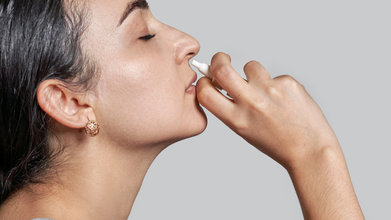
Nasal Spray Warning Over 'Rebound Congestion'; Experts Say It Should Not Be Used For More Than 7 Days
Credits: iStock
Nasal spray warnings are given by doctors and experts as the long use of it could lead to worsening of condition. The Royal Pharmaceutical Society (RPS) advised the public to not use nasal decongestant for more than seven days, as it contains xylometazoline or oxymetazoline. Its prolonged use can cause 'rebound congestion' or increased dependency on these sprays to breathe easily.
A recent poll, reports the Independent found out that almost six in 10 pharmacists report patients were unaware of the dangers of its extended use. Due to the increased number of flu activity, symptoms like blocked nose has increased, which has lead to a high usage of nasal spray.
Is Rebound Congestion Preventable?
It is a preventable condition, and is scientifically known as rhinitis medicamentosa, which causes the symptoms to worsen. Patients become depended on the sprays to breathe more easily.
RPS survey of 300 pharmacists found that 59% think the public is not aware of the risks, while 75% said packaging should be clearer about the seven-day limit. 63% said they had intervened in cases of suspected overuse.
Professor Amira Guirguis, chief scientist at RPS told the ITV News, "Nasal decongestant sprays can be helpful for short-term relief, but using them for longer than seven days can make your congestion significantly worse. Our research shows that many people are unaware of this risk, which means they may continue using these sprays without realizing they could be prolonging their symptoms. We'd like to see clearer warnings on the packaging which you can't miss and greater awareness of the seven-day limit. If your congestion lasts more than a week, speak to your pharmacist. There are safe and effective alternative options to help you manage your symptoms."
Another survey by ITV News suggests that more than a fifth of adults have used the products for longer than seven days. This means 5.5 million people in the UK may have risked developing a dependency.
Read: Bristol Hospitals Under Severe Strain as Flu and Cold Weather Hit the Region
As ITV News reported, Charlotte Johnstone, who is 30, had been using nasal spray multiple times a day since she was seven years old. She realized that the impact of this so-called addiction has caused her anxiety and left her "dreaming about not being able to breathe". She told ITV News, “I can’t sleep without having it, I wake up and the first thing I do is have my nasal spray. I don’t like eating if I’ve got a blocked up nose, it just makes me feel claustrophobic. I wouldn’t put myself in a situation where I don’t have it. “I go through stages of losing my sense of smell. I know it’s doing something but I don’t know what. But for the sake of having a clear nose, I’ll just take it,” she said.
A spokesperson from PAGB, the consumer healthcare association representing the manufacturers of these products, said: “The patient information leaflet which accompanies all nasal decongestant sprays, includes these instructions and outlines the risks of taking the medication for longer than its indicated use. As explained on the information supplied with the nasal decongestant sprays, OTC medicines manufacturers provide comprehensive accessible information to support people to make responsible informed decisions about the right product to self-care for their self-treatable condition," reported ITV News.
- Follow Us :


© 2024 Bennett, Coleman & Company Limited