- Health Conditions A-Z
- Health & Wellness
- Nutrition
- Fitness
- Health News
- Ayurveda
- Videos
- Medicine A-Z
- Parenting
- Web Stories
Global Premature Deaths Will Be Halved By 2050, Says Lancet Report

Just before the 2024 World Health Summit, of which the World Health Organisation (WHO) is a partner, the Lancet Commission on "Investing in Health" published a report with an "ambitious" yet "feasible" goal: cutting the global probability of premature deaths by 50% by the year 2050. The "50-by-50" initiative aims to ensure that a child born in 2050 has only 15% chance of dying before the age of 70, as compared to the children born in 2016, who are at 31% risk of dying before 70.
Global Leaders And Health Outcomes
Seven countries, including Bangladesh, Ethiopia, Iran and Türkiye, found the study, and were already on the track to achieve this goal. These countries have made progress in reducing premature death rates and are also now considered as model countries, serving examples for others who are aiming to improve universal health coverage.
The authors of this report are a team of researchers who come from prominent health institutions like Harvard University's School of Public Health, WHO and India's Public Health Foundation. They underscore that these countries' successes stem from early health interventions.
What Can Be Done To Reduce Early Deaths?
The authors have noted that expanding access to just 15 priority health services could make a great difference in reaching the 50-by-50 goal. These services address 8 maternal and infectious diseases and 7 non-communicable diseases (NCD) and injuries.
However, the biggest challenge remains the access to healthcare due to lack of funding and a gap in universal health coverage initiatives. "The most efficient route is to focus resources against a narrow set of conditions and scale up financing to develop new health technologies," noted the authors.
Prevention And Policy
The report points out tobacco control as one of the most effective and crucial measures to be carried out to reduce early deaths as it can reduce the tobacco-related deaths. There are policies being discussed to raise tax on tobacco that could turn out to be transformative.
Another aspect that could be taxed is unhealthy foods and beverages like sugary drinks since these are also linked to a higher risk of premature deaths, especially for those who have type 2 diabetes.
The Way Forward
The authors also outlined a target for a 30% reduction in premature deaths by 2035. The authors also emphasised that the economic impact of decreased mortality rates would also be substantial.
Breast Cancer Awareness Month: Nutritionist Reveals 6 Foods That May Lower Breast Cancer Risk

Credits: Canva
October is Breast Cancer Awareness Month, making it the perfect time to focus on habits that can help protect your health. While no single food can guarantee prevention, research shows that certain nutrients and antioxidants found in everyday foods may play a role in reducing the risk of breast cancer. For women, especially those with risk factors, incorporating these foods into your diet can be a meaningful step toward prevention.
Understanding Breast Cancer
Breast cancer occurs when breast cells start to grow abnormally and form tumours. If these tumours are not treated, they can spread to other parts of the body and become life-threatening. According to the World Health Organization (WHO), this disease claimed an estimated 670,000 lives worldwide in 2022. In India, breast cancer is also increasing, with one in 28 women now at risk. This makes it all the more important to understand ways to lower risk through lifestyle and dietary choices.
6 Foods To Prevent Breast Cancer
Nutritionist and weight loss specialist Leema Mahajan took to her Instagram to highlight the power of food in breast cancer prevention. She emphasizes starting early: “Introducing these foods into your diet from a young age can help reduce your chances of developing this disease later in life.” Mahajan identifies six key foods backed by research for their potential protective benefits.
1. Pomegranate
Pomegranates are rich in compounds called ellagitannins, which studies suggest may help slow down the growth of cancer cells and limit estrogen-driven tumour development. Mahajan recommends enjoying one cup of fresh pomegranate each day. “Fresh pomegranate seeds can support your body’s fight against abnormal cell growth. It’s simple, delicious, and effective,” she says.
2. Cruciferous Vegetables
Vegetables like broccoli, cauliflower, cabbage, and Brussels sprouts belong to the cruciferous family, known for their cancer-fighting properties. These vegetables contain sulforaphane, a compound that aids the liver in breaking down harmful estrogen by-products and may help prevent tumour formation. Mahajan advises, “Include cruciferous vegetables in your meals three to four times a week. You can eat them raw in salads or lightly steamed to retain their nutrients.”
3. Berries
Berries such as blueberries, strawberries, and raspberries are packed with antioxidants, particularly anthocyanins, which help combat cell damage and inflammation. Adding a handful of berries to your breakfast or as a snack is a tasty and nutritious way to support your body’s natural defenses.
4. Fatty Fish
Omega-3 fatty acids found in salmon, mackerel, and sardines may have anti-inflammatory properties that reduce cancer risk. Including fatty fish two to three times a week can support overall health and may help maintain hormone balance.
5. Green Tea
Green tea contains polyphenols, which have been shown to slow cancer cell growth in laboratory studies. Drinking two to three cups daily can be a simple, comforting habit with potential protective benefits.
6. Tomatoes
Tomatoes are rich in lycopene, an antioxidant linked to lower cancer risk. Cooking tomatoes, such as in sauces or soups, makes lycopene more easily absorbed by the body.
Small Changes, Big Impact
Incorporating these foods into your daily meals isn’t just about prevention, it’s also about building a sustainable, healthful lifestyle. While diet alone cannot prevent breast cancer, combining these nutritious foods with regular exercise, adequate sleep, and routine medical screenings can strengthen overall health and reduce long-term risk.
Eating with awareness, enjoying colorful fruits and vegetables, and making consistent, balanced choices can empower women to take charge of their health, one meal at a time.
Cancer Risk May Increase Depending On Where You Carry Fat: Study Links Excess Fat In This Part Of Body To Cancer

(Credit-Canva)
New research suggests that the common way doctors measure a healthy weight, called Body Mass Index (BMI), may not be the best tool for figuring out a person's risk for cancer. Instead, the study found that where a person carries their extra fat, especially fat around the stomach, is a much more important clue.
A new study published in the Journal of the National Cancer Institute, found that belly fat plays a big role, not just in our general health but also in cancer. However, how does fat around your mid-section define this?
Where Your Fat Sits May Be Key
For many years, doctors have used BMI (which is calculated from your height and weight) to see if someone is overweight or obese and then use that status to estimate their risk for various cancers. However, this new study found that relying only on BMI is too basic. The research suggests that the location of extra fat on your body might be just as important, if not more so, in deciding whether your risk of developing several types of cancer goes up or down.
How Belly Fat Increases Risk Of Cancer
The study used advanced methods to look at the relationship between fat in five different areas of the body and the risk for 12 common cancers linked to being overweight. The clearest and most alarming finding was that fat around the abdomen (belly fat) was the most dangerous type of fat:
Increased Risk
Belly fat was directly linked to a higher chance of developing three specific cancers: endometrial cancer (of the uterus), esophageal cancer (in the food pipe), and liver cancer.
Worse Health Marker
Doctors pointed out that carrying fat in the center of your body is already known to cause problems like diabetes and heart disease. They believe this is because central fat causes a lot of inflammation inside the body, and this inflammation is what can actually encourage cancer to grow.
Are There Benefits Of Having Belly Fat?
While fat around the stomach was dangerous, the researchers made a surprising discovery about lower body fat. They found that fat carried in the buttocks and thighs actually seemed to help protect against cancer. This fat was linked to a reduced risk of both breast cancer and a type of brain tumor called meningioma. This unexpected result has led some experts to think that the fat cells in the lower body might produce helpful hormones that fight against cancer.
Why BMI Isn't Enough
This research strongly confirms that doctors shouldn't just rely on the single BMI number. As one cancer doctor noted, BMI is a "quick and dirty" measurement that only gives a very basic picture of your health; the location of excess fat matters much more. Scientists believe fat increases cancer risk in a few ways, including:
- Producing extra hormones like estrogen and insulin, which are known to fuel the growth of many cancer types.
- Creating a state of chronic inflammation in the body, which helps tumors get started and grow.
The study concludes that future health plans, both for treating obesity and preventing cancer, should focus on measuring and managing where fat is distributed rather than just focusing on overall weight. While losing weight is still beneficial, the body part where the fat is concentrated is a powerful indicator of risk.
First Kidney Transplant With Universal Blood Type Performed: Here's How Scientists Achieved This
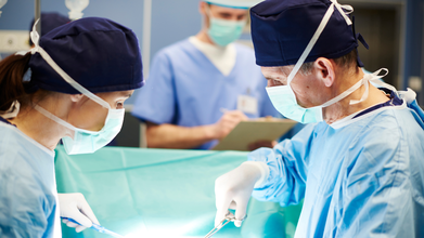
(Credit-Canva)
There are many aspects of an organ donation, not just the need for an organ, but also how compatible it is in terms of blood type. The reason why this is important is because of components known as antigens. The recipient's own body can destroy the new organ if the donated organ has different antigens. However, scientists may have found a way to surpass this issue.
A major scientific breakthrough has occurred: scientists successfully changed the blood type of a donated kidney using a special enzyme and then transplanted it into a patient. This is the first procedure of its kind. Right now, a patient can only receive an organ if the donor has a compatible blood type. If the types don't match, the patient's immune system will immediately produce antibodies or immune soldiers to attack and destroy the foreign organ.
How Did The Mismatched Blood Type Transplant Work?
The research team from Canada and China found a special enzyme that can remove the Type A antigens from an organ. Antigens are the markers on blood cells (and organs) that determine blood type. By removing the Type A markers, the enzyme essentially converts Type A blood into Type O blood.
Type O is considered the "universal" donor type because anyone can receive it. In this first test, the converted Type O kidney was transplanted into a 68-year-old patient. Although the kidney eventually showed signs of rejection after two days, it was able to function and produce urine for six days, proving the concept works.
How Does A Universal Kidney Work?
If doctors can successfully change the blood type of a donor organ, the current limits on transplants would largely disappear, creating enormous benefits for patients.
Significantly Increased Access to Organs
Without the need to match blood types, doctors wouldn't have to wait for an organ that is both a good match and the correct blood type. Instead, they could focus only on other factors important for the transplant's long-term success. By removing the blood type obstacle, this technology would allow for faster matching and quicker surgeries, which will dramatically reduce the painfully long waiting lists that patients currently face.
Shorter, More Efficient Wait Lists
Without the restriction of matching blood types, doctors wouldn't have to wait for an organ that is both a good match and the correct blood type. Instead, they could focus on other crucial factors, like matching proteins and tissues that are important for the long-term success of the transplant. By eliminating the blood type hurdle, this technology would allow for faster matching and quicker surgeries, drastically reducing the painfully long wait lists that patients currently face for life-saving organs.
Eliminating Risky Pre-Treatment for Recipients
This new approach of treating the donor organ instead of the patient is a major advancement. Right now, to perform a blood-type-mismatched transplant, the patient has to get intense treatments beforehand to severely weaken their immune system. These treatments are risky because they significantly increase the patient's vulnerability to severe infections.
Furthermore, for deceased donor organs—which must be used very quickly—there simply isn't enough time to safely administer these pre-treatments. By converting the organ outside the body, doctors can avoid this high-risk step for the recipient, making the transplant safer and much more feasible for emergency cases.
What Happens Next
This successful first attempt proves the enzyme-conversion technique works, but it's just the beginning. The scientists now need to conduct more studies on both brain-dead and living patients. Their main goal is to figure out the best way to adjust the treatment so that the converted organ can function successfully for a long period, ultimately allowing this revolutionary technique to be used in hospitals everywhere.
- Follow Us :


© 2024 Bennett, Coleman & Company Limited