- Health Conditions A-Z
- Health & Wellness
- Nutrition
- Fitness
- Health News
- Ayurveda
- Videos
- Medicine A-Z
- Parenting
What Really Happens in Hernia Surgery? A Clear, Patient-Friendly Guide to All the Types Explained
Credits: Canva
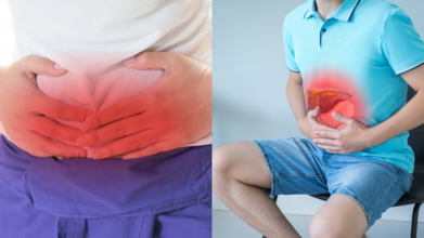
You bend to lift a bag, sneeze a little too hard, or notice a strange bulge that was not there before. That small lump in your abdomen could be a hernia. While the word itself can sound alarming, hernia surgery today is routine and safe and comes in different types depending on your condition.
What’s a hernia?
A hernia is when an organ, often the gut or fatty tissue, pokes through a weak spot in your abdominal wall. Dr Pushkar Anand Singh, Senior Consultant – General and Laparoscopic Surgeon at Shri Ram Singh Hospital, explains it like this: “Think of it like a bulge in a worn-out tyre. The wall is supposed to hold everything in, but a weak spot lets the insides push out.”
This weak spot may form due to overexertion, like unsupervised weightlifting, constipation, or chronic cough, or it could be congenital (you were born with it). Sometimes it even develops at the site of an old surgery.
Who actually needs surgery?
Not everyone who discovers a hernia needs to be wheeled into an operating theatre right away. According to Dr Shrey Srivastava, Senior Consultant in Internal Medicine at Sharda Hospital, Greater Noida, surgery is usually advised when the hernia becomes painful, interferes with daily activities, or risks dangerous complications.
“Hernias that cannot be pushed back in, those that cause increasing pain, or worse, strangulated hernias where blood supply is cut off, need urgent surgery,” he says. Without timely intervention, this can lead to tissue death and potentially life-threatening infection.
On the flip side, if a hernia is tiny, painless, and not causing trouble, your doctor may simply keep an eye on it. But the general rule is that a hernia will not magically disappear on its own; sooner or later, surgery might be on the cards.
Open Hernia Repair
This is the old-school, tried-and-tested method. Surgeons make an incision right over the hernia, carefully push the bulging tissue back into place, repair the defect, and then reinforce the area with a mesh (like patching up that worn-out tyre).
“Open repair is highly cost-effective, and the outcomes are comparable with newer techniques,” says Dr Singh. For many patients, it remains the go-to option, especially when budget is a concern.
Laparoscopic Hernia Repair
Here, instead of one big cut, surgeons make three or four tiny incisions to slip in a camera and surgical instruments. The mesh is placed from the inside, which means no obvious scar at the hernia site.
The perks are smaller cuts, quicker healing, and less pain afterwards. However, it costs more, and as Dr Singh points out, “It is not recommended for patients with significant heart or lung issues, as the procedure puts more strain on the body.”
Robotic-Assisted Hernia Repair
Robots in surgery may sound like something out of a sci-fi movie, but they are here. Robotic-assisted hernia repair builds on the laparoscopic method, except the tools are controlled through a robotic console.
“The technology allows greater precision and 3D visualisation for the surgeon,” explains Dr Singh. It is slick and advanced, but it also comes with a hefty price tag. Since the outcomes are not dramatically better than laparoscopic surgery, most hospitals do not see it as a routine option.
How do doctors decide which is right?
With three different techniques on the table, how do surgeons pick? Dr Singh says the choice depends on several factors like patient health, cost considerations, size of the hernia, and the urgency of the situation.
Open surgery might suit a patient looking for a straightforward, affordable solution. Laparoscopic repair works well for those who want a quicker recovery and can afford the added cost. Robotic surgery, while cool, is usually reserved for centres with access to the technology and patients willing to pay extra for the latest option.
Do not fear the word “surgery”
While the thought of going under the knife can be nerve-wracking, hernia surgeries today are routine, safe, and highly successful. Most patients return to normal activities within weeks, and the mesh reinforcement greatly reduces the risk of recurrence.
“Hernias are common, but complications can be dangerous,” Dr Srivastava reminds. “If you are experiencing persistent pain, visible bulges, or swelling that would not go back inside, do not delay consulting a doctor.”
UK Sees 11% Drop In Cancer Death Rates Over The Past Decade

Cancer deaths in the United Kingdom have dropped to their lowest recorded levels, according to new data from the charity Cancer Research UK. The figures show that cancer death rates have fallen by 11 per cent in the past decade, reflecting progress in early detection, screening, treatment and prevention.
Researchers estimate that around 247 people in every 100,000 in the UK now die from cancer each year. This is a significant decline from the peak recorded in 1989, when about 355 people per 100,000 died annually from the disease. Overall, that represents a 29 per cent reduction over the past few decades.
Experts say this steady improvement is the result of sustained scientific progress, improved healthcare systems and public health measures that target risk factors such as smoking.
Major Declines In Several Common Cancers
The new data highlights falling death rates across several major cancers. Ovarian cancer deaths have dropped by 19 per cent over the past ten years, up to 2024. Lung cancer deaths have fallen by 22 per cent during the same period, reflecting the long-term impact of reduced smoking rates and better treatment options.
Deaths from stomach cancer have seen one of the most dramatic improvements, dropping by 34 per cent in the past decade. Bowel cancer deaths have decreased by six per cent, while breast cancer deaths have fallen by 14 per cent.
Other cancers have also seen notable declines. Cervical and prostate cancer deaths have both dropped by 11 per cent. Deaths from leukemia are down by nine per cent, while esophageal cancer deaths have fallen by 12 per cent.
Experts say improved diagnosis, new therapies and better awareness are playing a key role in these trends.
Screening And Vaccination Making A Difference
One of the biggest public health successes has been the decline in cervical cancer deaths. Since the 1970s, deaths from cervical cancer have fallen by around 75 per cent in the UK. Health experts attribute much of this progress to the national cervical screening programme run by the NHS.
Screening helps detect abnormal cells early, allowing treatment before cancer develops or spreads.
Another major contributor is the human papillomavirus vaccine, commonly known as the HPV vaccine. The vaccine protects against the virus responsible for most cervical cancer cases. It is routinely offered to schoolchildren in the UK, and since its introduction in 2008, at least 6.5 million young people have received it.
Public health experts believe the vaccine will continue to reduce cervical cancer rates in the coming decades.
Some Cancer Death Rates Still Rising
Despite the overall progress, the data also shows worrying increases in deaths from certain cancers. Gallbladder cancer deaths have risen by 29 per cent, while deaths from eye cancer have increased by 26 per cent.
Liver cancer deaths are up by 14 per cent, and kidney cancer deaths have risen by five per cent. Meanwhile, death rates for thyroid cancer, pancreatic cancer and melanoma have remained largely unchanged.
Another important trend is that the total number of people dying from cancer continues to rise. This is largely due to population growth and an ageing population, as cancer risk increases with age.
Calls For More Research And Clinical Trials
Experts say the long-term decline in cancer deaths reflects decades of medical research and innovation. However, they stress that continued investment is necessary to sustain progress.
Cancer Research UK researcher Dr Sam Godfrey said the figures show the impact of scientific breakthroughs over many years. He has called on the government to support more clinical trials and ensure that NHS staff have enough time and resources to take part in life saving research.
Public health policies such as smoking bans, along with screening programmes and vaccines, are also credited with helping drive down cancer deaths across the country.
Scrolling Your Phone While In Toilet Can Spike Your Risk Of Hemorrhoids By 46%: Study

Credit: Canva
Are you in the habit of catching up on social media or news updates while sitting on the pot? A new study shows you may be "unintentionally" spending extra time and increasing your risk of developing painful hemorrhoids by 46 percent.
The study, published in the open-access journal PLOS One, explained that getting distracted by news or social media can increase pressure on sensitive anal tissues, which leads to hemorrhoids.
Hemorrhoids, also called piles, are swollen and inflamed veins around your anus or in your lower rectum.
"Using a smartphone while on the toilet was linked to a 46 percent increased chance of having hemorrhoids. We're still uncovering the many ways smartphones and our modern way of life impact our health,” Trisha Pasricha, from the Beth Israel Deaconess Medical Center in the US.
“It's possible that how and where we use them -- such as while in the bathroom -- can have unintended consequences," she added.
For the study, the researchers examined data from colonoscopies of 125 adults in America, and conducted an online survey to understand their lifestyle habits and behavior while using the toilet.
Two-thirds of the participants reported using their smartphones while on
the toilet. Compared with those who did not, endoscopists found that they had a 46 percent higher risk of hemorrhoids.
Longer Toilet Time And Mobile Scrolling
More than a third of bathroom smartphone users reported spending more than five minutes there during a single visit -- reading news (54.3 percent), or browsing social media (44.4 percent).
In comparison, just 7.1 percent of non-users reported staying that long.
"Smartphone use may unintentionally extend the time people spend sitting on the toilet. Sitting for longer periods could increase pressure on tissues in the anal region, which may contribute to the development of hemorrhoids,” the researchers said.
Pasricha suggested individuals leave smartphones outside the bathroom to understand the actual time it takes for a bowel movement.
"If it's taking longer, ask yourself why. Was it because having a bowel movement was really so difficult, or was it because my focus was elsewhere?" she said, calling for more studies.
Hemorrhoids: The Symptoms, Risk Factors
Studies estimate that hemorrhoidal disease affects 40% of people all over the world, and it is one of the most common diseases in the anorectal region.
The two types of hemorrhoids are:
- external hemorrhoids -- under the skin around the anus
- internal hemorrhoids -- in the lining of the anus and lower rectum
- bright red rectal bleeding during bowel movements,
- anal itching or irritation, pain or discomfort (especially while sitting),
- swelling or hard, tender lumps around the anus
- straining during bowel movements
- sitting on the toilet for too long
- chronic constipation or diarrhea
- eating low fiber foods
- older age >50
- pregnancy
- lifting heavy objects
- consume 25-35g of fiber daily,
- drink 6-8 glasses of water,
- avoid straining,
- limit toilet time to under five minutes
- do not delay bowel movements,
- maintain hygiene.
Black Rain Over Iran's Capital Tehran Sparks Health Fears

Credits: X/Twitter
Iran's capital Tehran was engulfed in a black cloud of toxic smoke. This also unleashed a black rainfall on Sunday after overnight Israeli strikes on several fuel depots caused fires to burn for hours. Images have come from across the city of Tehran. These images show thick black smoke from the fires hanging over it. Residents have also reported difficulty breathing and oil-tainted rainfall staining everything around them.
As per a TIME report, Iran's Red Crescent Society warned the residents of Tehran and the surrounding region that the rainfall after the strikes could be "highly dangerous and acidic", and could cause "chemical burns of the skin and serious damage to the lungs".
Many have complained about breathing problems, along with headache, feeling dry and sore lips, and feeling like burn in the eyes and constant itch in the throat.
What Does The Toxic Rain Fall Contain?
Iran's Red Crescent Society issued statements on Telegram that the rain could be contaminated with "toxic hydrocarbon compounds" as well as "sulfur and nitrogen oxides".
What Are The Health Risks?
As per a report by The Conversation, people exposed to the black smoke in Iran could experience headaches or difficulty in breathing, especially if they have asthma or a lung disease.
People who are more prone to health issues are older people, young children, anyone with disabilities and pregnant women. This could also lead to lower birth weights.
Since the thick black cloud from all the burning could increase the PM2.5 or the ultrafine particles, known as particulate matter, it could also increase cancer risks, along with neurological conditions and cardiovascular conditions.

The toxic rain could further pollute the natural waterways and drinking water sources. A photo shared by Iran's Red Crescent shows a healthcare worker's uniform covered in black droplets from the rain.
The "rain drops" are tainted with oily residue and could lead to skin problems, and if inhaled, it could also lead to serious medical crisis, noted Jim NR Dale, a senior meteorologist at British Weather Services.
It may also carry carcinogenic polycyclic aromatic hydrocarbons (PAHs) along with heavy metals that are released when construction materials burn and then remain suspended in the air.
As acidity increases, natural water bodies such as rivers and lakes can become too hostile to support life. When the pH of water drops below 5, most fish cannot survive, and at pH 4, a lake is often described as a “dead water body” because almost no living organisms remain.
Acid rain also harms the soil. It reduces calcium levels, an important nutrient for plants, and makes it easier for toxic aluminium to leach into water sources, further threatening ecosystems.
- Follow Us :


© 2024 Bennett, Coleman & Company Limited