- Health Conditions A-Z
- Health & Wellness
- Nutrition
- Fitness
- Health News
- Ayurveda
- Videos
- Medicine A-Z
- Parenting
- Web Stories
Polio Vaccination Campaign Begins In War-Torn Gaza, U.N. Stresses Success Only On A Pause In Fighting

As the conflict between Israel and Gaza continues to wreak havoc, a new threat has emerged in the region: polio. The ongoing war has exacerbated the already dire humanitarian situation in Gaza, leading to a resurgence of the poliovirus. In response, health officials have launched an emergency vaccination campaign to curb the spread of the virus.
Despite the challenges posed by the conflict, the campaign has brought a glimmer of hope to the beleaguered population.
A campaign to inoculate children in Gaza against polio and prevent the spread of the virus began on Saturday, the Health Ministry said, as Palestinians in the Hamas-governed enclave and in the occupied West Bank reeled from Israel's military offensives.
A small number of children in Gaza began receiving doses a day before the large-scale vaccine rollout and planned pause in fighting agreed to by Israel and the U.N. World Health Organization. The WHO confirmed the larger campaign would begin Sunday.
The polio outbreak in Gaza has raised alarms across the region, particularly as the virus threatens to spread beyond the enclave's borders. With the healthcare system already stretched thin due to the ongoing strikes and blockades, the situation has become increasingly precarious. The World Health Organization (WHO) and local health authorities have warned that without immediate intervention, the virus could lead to a public health crisis of unprecedented scale.
Amidst the conflict, Israel has agreed to a temporary pause in hostilities to allow for the safe distribution of vaccines. This truce, though fragile, has enabled health workers to begin administering the polio vaccine to children in Gaza, many of whom have been cut off from essential healthcare services for months.
“There must be a cease-fire so that the teams can reach everyone targeted by this campaign,” said Dr. Yousef Abu Al-Rish, Gaza's deputy health minister, describing scenes of sewage running through crowded tent camps.
Associated Press journalists saw about 10 children receiving doses at Nasser hospital in Khan Younis.
“I was terrified and waiting for the vaccination to arrive and for everyone to receive it,” said Amal Shaheen, whose daughter received a dose.
Israel is expected to pause some operations in Gaza on Sunday to allow health workers to administer vaccines with the aim of reaching some 640,000 Palestinian children. Officials said the pause would last at least nine hours and is unrelated to ongoing cease-fire negotiations.
The first batch of polio vaccines arrived in Gaza under tight security, with health workers racing against time to inoculate as many children as possible. The vaccination campaign is being spearheaded by the WHO, in coordination with the United Nations Relief and Works Agency (UNRWA) and the Palestinian Ministry of Health. The campaign targets children under the age of five, who are most vulnerable to the virus.
The three-day vaccination campaign comes after the first polio case in 25 years in Gaza was discovered this month. Doctors concluded a 10-month-old had been partially paralyzed by a mutated strain of the virus after not being vaccinated due to fighting.
Healthcare workers in Gaza have warned of the potential for a polio outbreak for months. The territory's humanitarian crisis has deepened during the war that broke out after Hamas-led militants stormed into southern Israel on Oct. 7, killing some 1,200 people and abducting around 250. Israel’s retaliatory offensive has killed more than 40,000 Palestinians, according to Gaza’s Health Ministry, which does not say how many were militants.
Despite the ceasefire, distributing the vaccine poses significant challenges. Gaza's healthcare infrastructure has been severely damaged by the ongoing conflict, and many areas remain inaccessible due to the destruction of roads and facilities. Moreover, the constant threat of renewed fighting looms large, making it difficult for health workers to reach certain areas. However, the international community has rallied to support the campaign, with additional vaccines and medical supplies being sent to Gaza.
The situation in Gaza remains volatile, with the possibility of renewed hostilities threatening to derail the vaccination campaign. Health officials have expressed concerns that the ceasefire may not hold long enough to complete the vaccination drive. Additionally, the psychological toll of the conflict on both health workers and the population at large cannot be underestimated. Many families are reluctant to bring their children to vaccination centers due to fear of airstrikes or violence.
Despite these challenges, there is hope that the vaccination campaign will succeed in curbing the spread of polio in Gaza. The WHO has stressed the importance of immunization in preventing a full-blown epidemic, and has called on all parties involved in the conflict to prioritize the health and well-being of the region's children. The international community has also pledged continued support, with promises of more vaccines and medical aid in the coming weeks.
The fragile truce has given Gaza a brief respite from the violence, allowing health workers to carry out their mission of protecting the most vulnerable. However, the situation remains precarious, and the success of the vaccination campaign hinges on the maintenance of the ceasefire and the continued flow of aid into the region.
Björn Borg Shares His Experience Of Living With Prostate Cancer

Credits: Wikimedia Commons
Björn Borg, the 11-time Grand Slam champion, a name the world is not unknown from has shocked everyone with his autobiography, where he revealed that he has been living with prostate cancer. His autobiography, Heartbeats is due for release on September 18, however, some of the details from his books were leaked on Thursday.
The details, which has been shared with The Athletic, Borg talks about his life-saving surgery that he had got done a couple years ago. He explained that "there never really was a choice because the cancer turned out to be extremely aggressive".
How Did Borg Find Out About His Prostate Cancer?
He reveals that his cancer was picked up because he would self examine regularly even though he did not have the symptoms. He revealed that the operation was necessary because "it turned out I was in the most advanced stage".
He said: "The fear comes and goes. But that's life, isn't it? You never really know what's coming. You just have to make the most of each day."
Also Read: Joe Biden Is Diagnosed With Aggressive Prostate Cancer: All That You Need To Know
What Is Prostate Cancer?
As per the National Cancer Institute, USA, prostate cancer is the most common cancer and the second leading cause of cancer death among men in the United States. Prostate cancer usually grows very slowly, this is why finding it and treating it before symptoms occur could improve men's health.
The National Health Service (NHS), UK, notes that this affects the prostate gland under the bladder and usually is common among men over 50 years old. The prostate is a small, walnut-size gland under the bladder, which produces a thick, white fluid that mixes with sperm to make semen.
How aggressive the cancer is depends on if it has spread to other parts of the body. However, early diagnosis could help it treat successfully.
Are There Any Common Symptoms Of Prostate Cancer?
The NHS, UK notes that prostate cancer usually do not have signs at first, but it could have certain symptoms later on. The best way is to get regular screenings done to identify early, especially if you are in your late 40s.
Also Read: Olympic Cyclist Sir Chris Hoy Shares His Diagnosis Of Prostate Cancer From A Common Shoulder Pain
Common Symptoms Of Prostate Cancer
As per the US Centers for Disease Control and Prevention (CDC), these are the symptoms common in prostate cancer. Speak to your doctor if you experience any such symptom:
Difficulty starting urination
Weak or interrupted flow of urine
Urinating often, especially at night
Trouble emptying the bladder completely
Pain or burning during urination
Blood in the urine or semen
Pain in the back, hips, or pelvis that doesn't go away
Painful ejaculation
Other Health Issues Borg's Book Covers
Another video interview from a couple days ago shows Borg excited about his book launch that he has written with his wife Patricia. He, in the interview, described many near-death experiences and even talked about his cocaine addiction after his early retirement from tennis in his mid-20s.
Read: Can Omega-3-rich Food Slow Prostate Cancer Growth?
However, he is now focused as he calls himself a "family man". He now lives in Stockholm, and regularly spends time in Ibiza. "I have two beautiful sons, Robin and Leo [a pro tennis player once ranked No. 334]. I have two beautiful grandchildren, aged 12 and 10. And I’m kind of a family man, and I want to spend a lot of time with the family. And that’s important for me. Stay involved with tennis, to travel, to watch tennis, to spend time with my tennis friends, with the people who love tennis, but to be with the family, with the kids, with the grandchildren, that’s very important to me."
First Symptom Of Alzheimer’s Is Triggered By This Signal
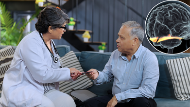
Credits: iStock
A fading sense of smell may do more than hint at aging—it could be one of the earliest warning signs of Alzheimer’s disease. New research from DZNE and Ludwig-Maximilians-Universität München reveals that immune cells in the brain mistakenly target and destroy nerve fibers critical for odor perception, offering fresh clues into how the disease begins and how it might be diagnosed sooner.
Patients and families may notice that scents seem muted or distorted, sometimes years before other symptoms. Until now, the exact cause behind this early warning sign remained elusive.
A new study led by researchers at the German Center for Neurodegenerative Diseases (DZNE) and Ludwig-Maximilians-Universität München (LMU) sheds fresh light on the mystery. The findings, published in Nature Communications, suggest that the brain’s own immune system mistakenly destroys nerve fibers vital for processing odors. This breakthrough could open the door to earlier diagnosis—and possibly earlier treatment—of Alzheimer’s.
How the Brain Processes Smell?
Smell perception begins in the olfactory bulb, a small but complex structure in the forebrain that receives input from sensory receptors in the nose. But the olfactory bulb doesn’t work alone. It relies on nerve fibers extending from the locus coeruleus, a brainstem region that helps regulate attention, blood flow, and sensory processing.
The new study shows that in early Alzheimer’s disease, this communication line is disrupted. Microglia—immune cells that normally act as the brain’s cleanup crew—start dismantling the nerve fibers linking the locus coeruleus and olfactory bulb. This immune-driven attack deprives the brain of crucial odor-processing pathways, leading to smell loss.
“Our study suggests that in early Alzheimer’s disease, changes occur in the nerve fibers linking the locus coeruleus to the olfactory bulb. These alterations signal to the microglia that affected fibers are defective or superfluous. Consequently, the microglia break them down,” explained Dr. Lars Paeger of DZNE and LMU.
What Is The “Eat-Me” Signal That Triggers Alzheimer’s?
At the heart of this immune misfire lies an unusual molecular change. Researchers observed that phosphatidylserine, a fatty acid normally tucked inside the protective membrane of neurons, shifts to the cell’s outer surface in affected nerve fibers.
When this happens, microglia interpret it as an “eat-me” signal. Under normal conditions, this signal supports a healthy process called synaptic pruning, where unnecessary or damaged connections are cleared away. But in Alzheimer’s, this mechanism seems to go awry.
Paeger explained that hyperactive neurons—cells firing abnormally due to early disease changes—appear to trigger this membrane shift. Once flagged as dysfunctional, these otherwise critical smell-related fibers are targeted and destroyed.
PET imaging scans in living patients, which confirmed damage to smell-related nerve circuits in people with Alzheimer’s or mild cognitive impairment.
“Smell issues in Alzheimer’s disease and damage to the associated nerves have been discussed for some time. However, the causes were unclear until now. Our findings point to an immunological mechanism as cause for such dysfunctions – and, in particular, that such events already arise in the early stages of Alzheimer’s disease,” said Prof. Joachim Herms, senior researcher on the study.
Why Smell Loss Is Important in Diagnosis of Alzheimer’s?
More than 6 million Americans currently live with Alzheimer’s disease, a number projected to rise sharply as populations age. Yet diagnosis often comes late, when memory loss and cognitive decline are already advanced.
Smell loss offers an earlier red flag. If the biological mechanisms behind it can be mapped and measured, doctors could potentially screen for Alzheimer’s years before symptoms interfere with daily life. This could be especially critical as new treatments, such as amyloid-beta antibodies, are designed to work best in the earliest phases of disease progression.
“Our findings could pave the way for the early identification of patients at risk of developing Alzheimer’s, enabling them to undergo comprehensive testing to confirm the diagnosis before cognitive problems arise,” Herms noted. “This would allow earlier intervention with amyloid-beta antibodies, increasing the probability of a positive response.”
The study also highlights the often-overlooked locus coeruleus, a small cluster of neurons deep in the brainstem. Beyond smell, this structure regulates blood flow, sleep-wake cycles, and stress responses through widespread connections.
Damage to the locus coeruleus is one of the earliest detectable signs of Alzheimer’s, even before amyloid plaques and tau tangles spread widely. The new findings underscore that its breakdown may not only affect memory and attention but also disrupt sensory pathways, making smell loss one of the first noticeable symptoms.
Can Sense of Smell Be Preserved?
While the research answers critical questions, it also raises new ones. If immune cells misidentify smell-related fibers as expendable, can this process be slowed or stopped? Could therapies aimed at stabilizing neuron membranes prevent the fatal “eat-me” signal from being displayed in the first place?
Future studies will likely explore whether drugs that regulate microglial activity—or protect the integrity of phosphatidylserine positioning—could preserve nerve connections in early Alzheimer’s.
Meanwhile, incorporating smell testing into routine checkups for older adults may become a more practical step. Inexpensive scratch-and-sniff exams already exist and could serve as a noninvasive screening tool to identify people who may need further evaluation.
For families watching loved ones struggle with subtle changes—whether misplaced keys, unusual forgetfulness, or an inability to smell morning coffee—the findings offer clarity. Smell loss is not just an odd, isolated symptom but a biological signal that Alzheimer’s is affecting the brain long before dementia sets in.
Recognizing this signal early could give patients and clinicians a window of opportunity: time to prepare, time to plan, and perhaps in the future, time to intervene with treatments that slow or alter the course of disease.
Alzheimer’s remains one of the greatest public health challenges of our time. While there is still no cure, understanding its earliest signals brings us closer to meaningful change. By uncovering how immune cells mistakenly dismantle smell pathways, this study not only solves a long-standing puzzle but also lays the foundation for earlier, more effective care strategies.
The sense of smell may be more than just a window to the world—it may be a window into the earliest changes of Alzheimer’s disease.
Tiny 3D-Printed Spinal Cords Could Reverse Paralysis, How Did Scientists Make It Work?
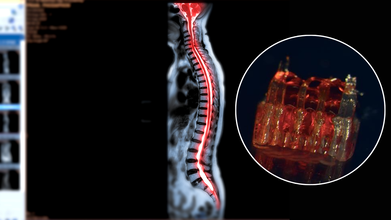
Credits: Canva/McAlpine Research Group, University of Minnesota
Spinal cord injuries have long posed one of the most stubborn challenges in medicine. Affecting more than 300,000 people in the United States alone, these injuries often lead to permanent paralysis because damaged nerve fibers fail to regenerate across the site of trauma. Traditional therapies focus largely on rehabilitation and symptom management rather than reversing the underlying injury. Now, a groundbreaking study from the University of Minnesota Twin Cities suggests that a combination of 3D printing, stem cells, and lab-grown tissues could change that narrative. Researchers have engineered tiny scaffolds that guide stem cells to form nerve fibers capable of bridging severed spinal cords. In rat models, this approach restored nerve connections and movement—offering a tantalizing glimpse into the future of paralysis treatment.
At the heart of this innovation are organoid scaffolds—microscopic 3D-printed structures designed to direct stem cell growth. These scaffolds contain a network of tiny channels that can be seeded with spinal neural progenitor cells (sNPCs). Originating from human adult stem cells, sNPCs have the potential to differentiate into the various types of neurons needed for spinal cord repair. The scaffold essentially provides a framework for these cells, ensuring they grow along the correct pathways to reconnect disrupted nerve circuits.
Guebum Han, a former postdoctoral researcher in mechanical engineering at the University of Minnesota and the study’s first author, explains, “We use the 3D printed channels of the scaffold to direct the growth of the stem cells, which ensures the new nerve fibers grow in the desired way. This method creates a relay system that, when placed in the spinal cord, bypasses the damaged area.”
To test their approach, the researchers transplanted the scaffolds into rats with completely severed spinal cords. Over time, the stem cells differentiated into mature neurons and extended their nerve fibers in both directions—toward the head (rostral) and toward the tail (caudal)—forming new connections with the host’s existing spinal circuitry.
The results were remarkable. Rats that received the organoid scaffolds showed significant functional recovery compared to controls, regaining movements that were previously impossible. The new neurons integrated seamlessly into the host tissue, demonstrating that lab-grown spinal tissue could not only survive transplantation but also restore communication across previously severed areas.
Can Lab-Grown Organs Help Patients?
While the research is still in its early stages, the potential implications for human medicine are profound. Spinal cord injuries have been notoriously resistant to treatment because adult nerve cells rarely regrow once damaged. This study provides proof-of-concept that targeted, scaffold-guided stem cell growth can rebuild the neural network necessary for motor function.
Ann Parr, professor of neurosurgery at the University of Minnesota, emphasizes the significance: “Regenerative medicine has brought about a new era in spinal cord injury research. Our laboratory is excited to explore the future potential of our ‘mini spinal cords’ for clinical translation.” The team hopes to refine the technique, scale up scaffold production, and move toward clinical trials that could one day benefit people living with paralysis.
Despite the promising results, several hurdles remain before this technology can be applied to humans. Scaling the tiny lab-grown spinal cords to the size necessary for human injuries will require sophisticated bioengineering solutions. Immune rejection and integration into a complex, pre-existing nervous system present additional challenges. Moreover, safety and efficacy will need to be rigorously tested in larger animal models before human trials can proceed.
The ethical considerations of stem cell use and genetic manipulation also require careful navigation. While adult stem cells used in this study bypass some of the ethical debates associated with embryonic stem cells, clinical applications must still adhere to stringent regulatory standards.
The merging of 3D printing, stem cell science, and lab-grown tissue engineering represents a paradigm shift in regenerative medicine. The concept of “mini spinal cords” could open the door to therapies not only for spinal cord injuries but potentially for other neurodegenerative diseases that involve nerve degeneration, such as amyotrophic lateral sclerosis (ALS) or multiple sclerosis.
Moreover, these technologies exemplify the broader trend of personalized medicine. By tailoring organoid scaffolds to individual patients, it may become possible to repair nervous system injuries with unprecedented precision. This could drastically improve outcomes, reduce rehabilitation times, and enhance quality of life for patients who currently have few options.
The University of Minnesota study is an early but significant step toward reversing paralysis. By combining 3D-printed scaffolds, stem cell biology, and lab-grown spinal tissue, researchers have demonstrated that damaged neural pathways can be rebuilt and functional recovery is achievable—at least in animal models.
While human applications are still a way off, the research provides a blueprint for the future of spinal cord repair and regenerative neuroscience. For the millions affected by spinal cord injuries, these tiny lab-grown spinal cords could one day offer more than hope—they could offer a pathway to regained movement and independence.
- Follow Us :


© 2024 Bennett, Coleman & Company Limited