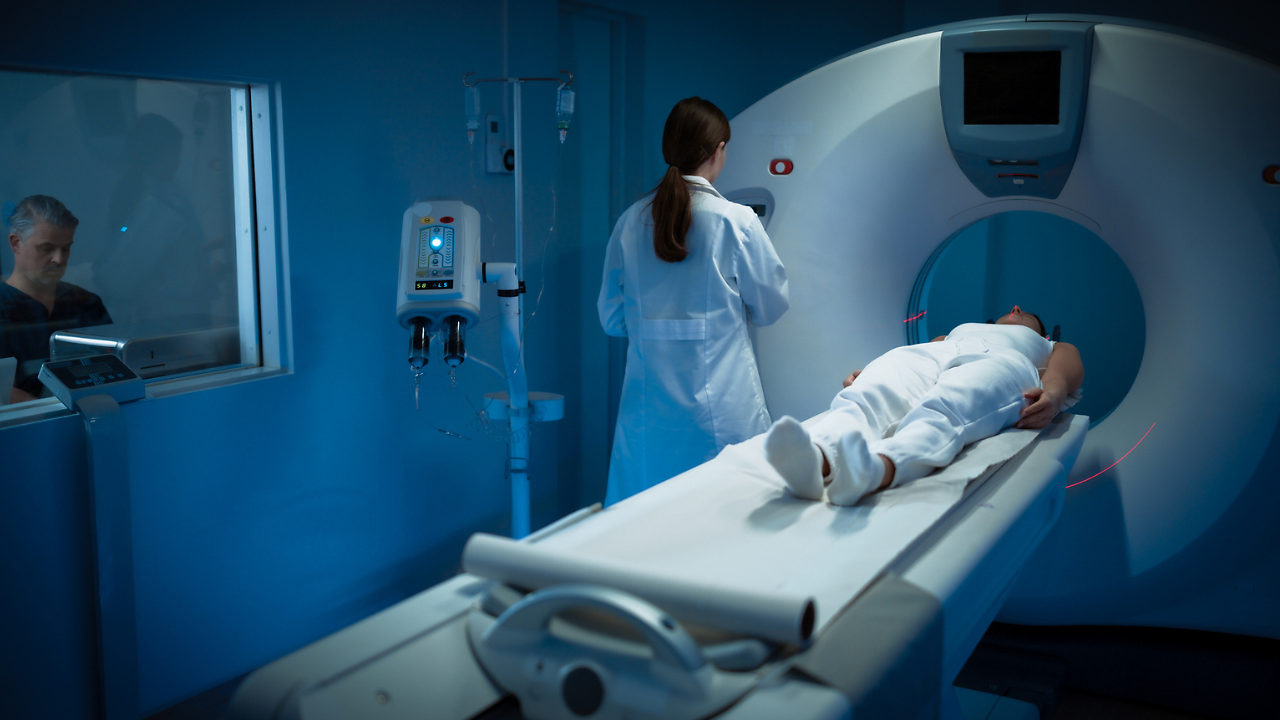
If you or someone you love has been diagnosed with colon cancer, chances are a doctor has already told you to go for a CT scan, maybe an MRI, and possibly mentioned a "PET-CT scan" at some point. It’s a valid question: Why do I need another scan, and what additional information can it provide that previous scans may not have shown? The short answer is that PET-CT isn't a routine test for colon cancer, and that's actually by design. It's a specialised tool that shines in a handful of specific situations, while adding little (and sometimes confusing) in others. Understanding where it fits can help you have a more informed conversation with your oncology team. What PET-CT Actually Shows A standard CT scan is like a detailed photograph of your insides; it shows the size, shape, and location of organs, tumours, and lymph nodes. A PET (positron emission tomography) scan works differently. Patients are injected with a small amount of a radioactive sugar tracer, most commonly FDG (fluorodeoxyglucose). Cancer cells tend to be metabolically hyperactive, so they take up more sugar than normal tissue, which shows up as a "hot spot" on the scan. A PET-CT combines both technologies in a single machine, overlaying the metabolic activity from the PET onto the precise anatomy from the CT. The result is a picture that shows not just where something is, but how biologically active it appears to be. Where PET-CT Genuinely Adds Value 1. Sorting Out Equivocal Findings on CT or MRI Sometimes a routine CT or MRI turns up an ambiguous spot, a small lung nodule, a liver lesion that doesn't clearly appear benign or malignant, or an enlarged lymph node of uncertain significance. In these cases, PET-CT can help clarify whether that finding is likely to be cancer-related or an incidental, harmless abnormality, potentially sparing a patient an unnecessary biopsy or, conversely, prompting one that's genuinely needed.Also read: The Neck Lump Most People Dismiss: When Should You Worry About Thyroid Cancer? 2. Working Up a Suspected Recurrence This is arguably where PET-CT earns its keep the most. After treatment, patients are typically monitored with periodic blood tests for a tumour marker called CEA (carcinoembryonic antigen), alongside imaging. If CEA levels start climbing but a conventional CT scan looks clean, that's a classic scenario for PET-CT. Because recurrent tumour deposits may become metabolically active before they are large enough to be clearly detected on standard imaging, PET-CT can sometimes identify recurrent disease that may not be detected on CT alone. A review of multiple studies has found that FDG-PET/CT can be highly sensitive for detecting recurrent disease when CEA levels are rising, but conventional imaging remains inconclusive. However, it is not perfect, and false-positive results can occur. 3. Pre-Surgical Planning Before Metastasectomy For a subset of patients whose cancer has spread to the liver or lungs, surgery to remove those metastases (sometimes combined with liver-directed therapies) can be curative or at least significantly life-extending. Before committing to a major operation like this, surgeons often want the most complete possible picture of disease extent. PET-CT is frequently used here to check for additional sites of disease outside the liver or lungs that might change the surgical plan, because there's little point in a complex resection if cancer has quietly spread elsewhere.Also read: India Caps Cancer Drug Trade Margins At 30%; Patients May Save Rs 2,500 Crore Annually 4. Resolving Discrepant or Confusing Results Occasionally, different scans or a scan and a physical exam simply don't agree with each other, or a biopsy comes back inconclusive. PET-CT can serve as a tiebreaker of sorts, helping the care team decide whether further investigation is warranted. Where PET-CT Is Not Recommended It's just as important to know when PET-CT doesn't help, because ordering it in the wrong setting can create more problems than it solves: false positives that trigger unnecessary biopsies, added cost, and added radiation exposure without a meaningful benefit. Initial staging. For most newly diagnosed colon cancers, standard CT of the chest, abdomen, and pelvis remains the primary staging tool. PET-CT is not routinely recommended upfront unless there's a specific unresolved question that CT can't answer. Routine surveillance after treatment. This is a common point of confusion. It might seem logical to add PET-CT to regular post-treatment monitoring to catch recurrence as early as possible. But a randomised trial comparing standard surveillance alone to standard surveillance plus routine PET-CT found no meaningful difference in recurrence detection between the two approaches. Major guideline bodies do not recommend routine PET-CT as part of standard surveillance protocols for exactly this reason; the added scans didn't translate into a clear benefit for patients. Monitoring response in metastatic disease already on treatment. For patients with established metastatic colon cancer undergoing systemic therapy, PET-CT isn't generally used as a routine tool to track how well treatment is working; CT remains the standard for that purpose.PET-CT is a precision tool, not a routine one. It tends to help most when there’s a specific, unanswered question, an ambiguous finding, a suspicious CEA rise despite clean conventional scans, or for pre-surgical planning before removing metastatic disease. Outside of these situations, standard CT and MRI generally remain the imaging workhorses for colon cancer, and PET-CT is not routinely recommended to detect recurrence earlier or monitor treatment response. If your care team is considering a PET-CT, it’s entirely reasonable to ask: “What specific question is this scan meant to answer?”By Dr. Digish Shah, Senior Consultant, Nuclear Medicine, KD Cancer Centre