- Health Conditions A-Z
- Health & Wellness
- Nutrition
- Fitness
- Health News
- Ayurveda
- Videos
- Medicine A-Z
- Parenting
From Anemia To Cancer: The Life-Threatening Illness No One Saw Coming

Credits: Canva
For weeks, Ann Malik, a 39-year-old mother of three and co-founder of two sports-related businesses, felt unwell but couldn’t identify the cause. With no significant medical history apart from mild asthma, she initially dismissed her fatigue and unease. When her doctor diagnosed her with mild anemia, she felt reassured and expected to recover with iron supplements, reports the Washington Post.
However, two months later, her condition worsened. She began experiencing anxiety, insomnia, loss of appetite, and weight loss. Suspecting depression, her physician prescribed an antidepressant, but it brought no relief. Malik started questioning why she remained unwell despite no apparent serious health concerns.
The Growing Concern
As her symptoms persisted, Malik found herself overwhelmed by an undefined but growing dread. She noticed a persistent fullness in the upper right side of her abdomen, though it wasn’t painful. Desperate for answers, she consulted an endocrinologist, suspecting a metabolic disorder, but blood tests showed nothing abnormal. The specialist attributed her symptoms to stress and suggested lifestyle changes.
Seeking alternative solutions, Malik consulted an integrative medicine practitioner who diagnosed her with “adrenal fatigue,” a controversial, non-medically recognized condition. She was advised to manage stress, adjust her diet, and take special serums. Though she tried to follow the recommendations, her health continued to decline. By May, she was experiencing severe night sweats, continued weight loss, and a persistent sense that something was terribly wrong.
A Misleading Diagnosis
In July, Malik developed a lingering cold that led to her coughing up blood. A chest X-ray revealed pneumonia in her right lung. She was prescribed antibiotics, which provided only slight relief. A month later, she coughed up blood again, prompting an emergency room visit. Another X-ray confirmed recurrent pneumonia, but her husband insisted something more serious was at play. They pushed for further testing, leading them to a pulmonologist.
A CT scan in August revealed an unusual area in her right lung. A bronchoscopy followed, allowing doctors to examine her airways and collect tissue samples. The results were shocking: Malik had non-small cell adenocarcinoma, the most common type of lung cancer.
A Devastating Diagnosis
Malik was stunned. A non-smoker with no prior indications of lung disease, she never suspected cancer. Her pulmonologist reassured her that they had hopefully caught it early. But Malik knew her symptoms had persisted for too long for this to be an early-stage diagnosis. The unexplained fullness in her abdomen now had an explanation—it was a sign of advanced cancer.
A Life-Threatening Turn
The day after her diagnosis, Malik’s health took a drastic turn. While attending her son’s preschool orientation, she developed double vision and struggled to use her hands. Assuming stress was to blame, she tried to push through. However, paramedics were called, and upon arrival at the hospital, doctors discovered she had suffered a stroke.
Further tests revealed the true extent of her illness. The lung cancer had spread extensively—to her left lung, liver, spine, hip bones, and brain. The stroke was a direct result of the cancer’s progression. Her prognosis was grim: doctors estimated she had about a month to live.
A New Hope: Genetic Testing and Targeted Therapy
Despite the dire outlook, Malik’s medical team pursued additional testing. Given her age and non-smoking history, they suspected her cancer might be linked to a genetic mutation. A specialized test revealed that she had a ROS1 mutation, a rare genetic alteration found in 1–2% of lung cancer patients, typically younger individuals who never smoked.
This discovery was crucial. ROS1-positive cancers respond to targeted therapy, a specialized treatment that attacks cancer cells with the specific mutation while sparing healthy cells. Malik began chemotherapy, and the treatment showed positive effects. She sought further expertise at Massachusetts General Hospital, where thoracic oncologists tailored a treatment plan for her unique condition.
A Remarkable Survival Story
Contrary to the initial prognosis, Malik defied the odds. Now, a decade after her diagnosis, she looks forward to celebrating her 50th birthday. Her case underscores the importance of patient advocacy, persistence, and advancements in genetic testing for precision medicine.
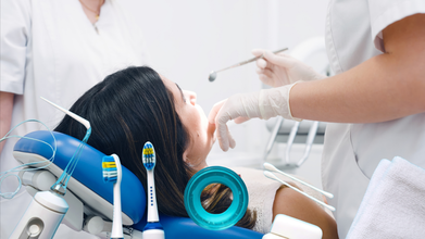
World Oral Health Day: Your Mouth May Signal Disease Before You Even Know, According To Doctor
Credits: Canva
Every year on March 20, World Oral Health Day is observed to raise awareness about the importance of caring for your mouth at every stage of life. This means starting from birth through later years. However, a visit to dentist is usually the last thing anyone plans when they think of a doctor's visit. Dental visits happen only when extraction or something bigger is involved. More often than not people ignore a follow up or a regular dental checkup to maintain oral health. Why so?
In a previous interview with Health and Me Lt Gen Dr Vimal Arora, the Chief Clinical officer at Clove Dental, who has 40 years of experience as a dentist and also served in the Indian Army explained how oral health is not just about a pretty smile. He explained that many do not plan a regular visit to a dentist because the problems that pertains to oral health do not seem "life threatening".
Read: Exclusive: Astronaut Rakesh Sharma Had To Get His Wisdom Tooth Extracted Before His Trip To Space
However, Dr Arora told Health and Me that oral health can in fact be the first way to know if anyone is prone to a chronic disease.
Early Signs Of Disease Your Mouth Can Reveal
“There is now scientific evidence, published in top medical journals, that shows how oral infections can significantly worsen pre-existing systemic diseases,” says Dr. Arora. This isn’t just a theory — it’s a medically established fact.
Take diabetes, for instance. People with gum infections often find it harder to control their blood sugar levels. “If you're suffering from diabetes and you have a gum infection, your diabetes will not be well controlled. In fact, it may worsen,” he adds. The same applies to heart diseases. Oral infections can increase inflammation in the body, potentially triggering or worsening cardiovascular problems.
This is concerning especially for those people who may already be managing conditions like hypertensions, arthritis, or respiratory illness. Poor oral health can actually sabotage their efforts to stay stable.
“Your tongue is a true mirror of your oral and general health,” says Dr. Arora. The correlation therefore goes beyond just gums and teeth. Dentists are trained to detect early signs of systemic disorders simply by examining the tongue's color, coating and texture.
A pale tongue might indicate iron deficiency, while a coated tongue could be a sign of digestive issues or a viral illness. “Sometimes, we ask patients if they've had their blood work done, based purely on what we see on their tongue,” he adds. In some cases, dentists can even detect undiagnosed diabetes or early signs of nutritional deficiencies.
What Mother's Oral Health Can Predict About Their Babies
Dr. Arora also highlights the overlooked risks that pregnant women face when they neglect their dental hygiene. “If you are pregnant and suffer from periodontitis or even gingivitis — essentially poor gum health — you are at risk of having a preterm or low-birth-weight baby,” he says.
This isn’t just theory; it’s a well-documented risk in obstetric dentistry. Pregnant women are encouraged to get dental check-ups not just for themselves, but for the health of their unborn child.
Despite these evidences, oral health remains one of the most neglected aspects of personal healthcare. Dr. Arora believes this is partly due to a lack of awareness and the perception that dental problems are not "serious enough."
“People often wait until they’re in pain before they see a dentist, but by then, the damage might already be affecting other parts of the body,” he says. Preventive check-ups, regular cleaning, and treating gum infections early on can go a long way in improving not just oral health, but overall wellness.
Fact Check: Sunscreen Is Only Used For Outdoors

Credits: Canva
Most people reach for sunscreen when heading to the beach or stepping out for a long day in the sun. But skincare experts say sun protection should not be limited to vacations or outdoor activities. Sunscreen is meant to be part of a daily skincare routine.
This often raises a common question. If you are staying indoors all day, do you still need sunscreen?
Dermatologists say the answer depends on your surroundings and daily habits.
Sunlight can still reach you indoors
Many people assume that staying inside completely protects their skin from sunlight. But this is not always true.
Dr Khushboo Jha, MBBS, MD, Chief Dermatologist Consultant at Metro Hospital and Founder of One Skin Clinic, explains that sunlight entering through windows can still affect the skin.
“While standard window glass blocks most UVB rays, which cause sunburn, UVA rays can still pass through. These rays penetrate deeper into the skin and are linked to long term concerns such as premature aging, uneven pigmentation and loss of skin elasticity,” she says.
These UVA rays are often overlooked because they do not cause immediate redness or burning like UVB rays. However, over time they can lead to visible signs of skin aging and pigmentation.
When sunscreen indoors becomes important
Dermatologists say sunscreen indoors is particularly useful for people who spend long hours near windows or in well lit spaces.
Dr Jha notes that individuals who work near windows, sit in sunlit rooms or spend time driving during the day may still be exposed to sunlight. “Even short periods of daily exposure to sunlight over time can contribute to cumulative skin damage,” she explains.
In such situations, applying sunscreen in the morning can offer an added layer of protection. A broad spectrum sunscreen with at least SPF 30 is generally recommended.
This approach is especially relevant for people working in offices with large windows or those who frequently commute during daylight hours.
What if you are away from windows?
Experts also say sunscreen use indoors is not always equally necessary for everyone.
If you spend most of the day inside a room with minimal natural light and away from windows, your exposure to ultraviolet radiation becomes much lower.
Dr Jha says that in such cases the urgency of frequent sunscreen reapplication becomes less important. The risk of sun related skin damage is significantly reduced when there is little to no direct daylight entering the space.
This means sunscreen indoors should not be treated as a strict rule but rather as a flexible part of skincare based on lifestyle and environment.
What about blue light from screens?
Another topic that often comes up is blue light exposure from digital devices such as phones, laptops and tablets.
Some studies suggest that prolonged exposure to visible light may contribute to pigmentation, especially in individuals with deeper skin tones. However, dermatologists point out that the amount of blue light from electronic screens is much lower than what we receive from natural sunlight.
Dr Jha says the effect of digital screens on the skin is still being studied, but compared to sun exposure, the impact remains minimal.
A practical approach to daily skincare
Dermatologists suggest viewing sunscreen as a preventive skincare habit rather than a rigid rule.
Dr Jha recommends incorporating sunscreen into your morning routine, particularly if your day includes stepping outdoors or spending time in naturally lit environments.
In simple terms, if daylight reaches your workspace or you plan to go outside later in the day, applying sunscreen in the morning is a small step that can help protect your skin over time.
Woman Left Medically Infertile After Seven Surgeries For Endometriosis That She Did Not Have

Credits: ABC News' Four Corners
At the age of 28, Courtney Paton realized she could never have children. She was medically infertile. This was after years of repeated surgeries, a total of seven, for 'suspected' endometriosis, due to which Dr Simon Gordon, Melbourne-based gynecologist removed both her ovaries and eventually her uterus.
Also Read: Menopause Can Raise Alzheimer Risk In Women, Neurologist Warns
Her story came to light through an investigation by the Australian Broadcasting Company or ABC's Four Corners, an investigation that looked at the treatment she received from Dr Gordon.
Courtney says she trusted the doctor completely. Now she says that trust has been shattered. “I feel completely betrayed by not only Simon Gordon, but by Epworth, by the healthcare system,” she told the program.
A Wrong Diagnosis That Led to Repeated Surgeries
Courtney first had laparoscopic surgery in 2018 with another surgeon, which confirmed she had endometriosis. The condition affects about one in seven Australian women and can cause severe pelvic pain and fertility problems.
Still struggling with pain, she began seeing Gordon in 2019 when she was 21.
Over the next several years she underwent seven surgeries with him. Gordon told her the procedures were necessary to treat severe endometriosis. Courtney and her family paid more than 32,000 Australian dollars for these surgeries alone.
But when investigators asked her to obtain her pathology reports, the results told a very different story. The tissue tests from most of her surgeries showed no evidence of endometriosis.
Despite this, operation reports written by Gordon continued to describe findings consistent with the disease.
Read: A Woman Lost Her Ovary To Endometriosis Surgery After Receiving An Ultimatum From Gynecologist
Removal of Both Ovaries
In 2021 Gordon removed one of Courtney’s ovaries, saying it was stuck to the pelvic wall. Later he removed the second ovary as well.
Independent specialists who reviewed the pathology for the investigation said the ovary appeared normal and there was no clear justification for removing it. One expert described the treatment as “unbelievable” after reviewing the medical records.
Medical guidelines generally advise caution when removing ovaries from young women who may want children in the future.
A Hysterectomy At 25
Despite losing both ovaries, Courtney continued to experience pelvic pain. Gordon later advised that she should undergo a hysterectomy.
Concerned, she sought a second opinion from another gynecologist who said the procedure was unnecessary and suggested non surgical treatments.
But after years of pain and repeated surgeries, Courtney says she felt desperate for relief and trusted the doctor who had treated her for so long. Her uterus was removed in 2023 when she was just 25.
Again, pathology results found no evidence of endometriosis.
Investigation and Legal Action
Courtney is now pursuing legal action through a medical negligence claim. The case has also drawn attention from regulators, with investigations underway into Gordon’s conduct.
Australia’s federal health minister Mark Butler described the allegations as “physically sickening”.
For Courtney, the emotional impact remains overwhelming.
“No woman should ever have to endure what I’ve endured,” she said. “I’ve had the opportunity to have a family taken away from me.”
- Follow Us :


© 2024 Bennett, Coleman & Company Limited