New Study Finds Plastics From Nanomedicines Could Be Transferred To Embryos

New Study Finds Plastics From Nanomedicines Could Be Transferred To Embryos, Image Credit - Unsplash

Text

Updated Jul 25, 2024 | 06:11 PM IST

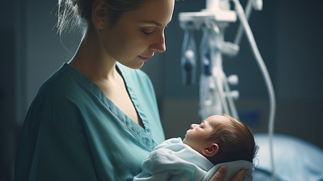
SummaryA recent study looked at the effects of nanomedicines on the embryo of a chicken test subject. They found microplastics in the baby. Find out what the implications of the study suggest

A new study published in Environment International has revealed Nanoplastics can still build up in newborn hearts that shed a new light on how such small plastic particle can create such a major hazard in our physical health.

What Are Nanoplastics?

It’s no secret that plastic has become an overabundant material in our daily use, from the cups we use at cafes to the packaging material for every small gift. Plastics that have been exposed to nature in particular start shedding small ‘nanoplastic’ particles wherever they go. Now, these tiny particles are posing a major problem.

From the oceans where we get our fish, to the soil where our plants are being uprooted to our very blood, nanoplastics have gotten anywhere. The presence of these particles in the human body pose a particular concern, as the researchers started to ask the question: what happens now if these nanoplastics are present inside newborns of the coming generation.

How Was The Study Conducted?

The study was conducted on chicken embryos by a group of researchers from Leiden University. According to the researchers, the study was conducted on chicken embryos, as it is easier to inject substances and take precise, observable measurements to see how the embryo develops inside the mother’s womb.

Since these nanoparticles are so small, it’s practically impossible to see them through conventional microscopes. As a result, the progress is tracked by tagging the nanoparticles with a clearly visible metal.

Their main finding was that these nanoplastics are capable of crossing the walls of the blood vessels themselves, leading them to build up to relatively high levels in the hart and kidneys.

Interestingly enough, the researchers also found nanoplastics in the avascular heart cushions that do not contain blood vessels. This suggests that these nanoplastics could pass into the heart of the embryo, directly through the fenestrate, which is a small opening that plays a role in forming and remodeling the heart’s structure during its development.

The study could potentially lead interesting results for the future, as we better our understanding of how these nanoplastics are spread to such an extent. Even if the outlook isn’t necessarily good, it could bring a change to how we approach our plastic consumption. As of right now, there’s already an understanding of how there needs to be more mindful use of nanomedicines when it comes to pregnant women.

End of Article

New Study Shows Dramatic Improvements In Heart Failure Treatment Across US Hospitals

New Study Shows Dramatic Improvements In Heart Failure Treatment Across US Hospitals

Credit: Canva

Text

Updated May 4, 2025 | 08:29 AM IST

SummaryLaunched in 2021, the American Heart Association’s three-year quality improvement initiative was designed to drive the uptake of quadruple medical therapy.

Heart failure continues to pose a major health threat in the United States, with approximately 6.7 million adults currently living with the condition. Alarming projections suggest that this number could climb to more than 8 million by 2030.

However, a promising new analysis published in Circulation: Heart Failure reveals that hospitals involved in the American Heart Association’s multiregional IMPLEMENT-HF initiative have significantly improved their adherence to guideline-directed medical therapy for patients with heart failure with reduced ejection fraction (HFrEF)—the most prevalent form of heart failure.

Launched in 2021, the American Heart Association’s three-year quality improvement initiative was designed to drive the uptake of quadruple medical therapy and incorporate assessments of patients’ health-related social needs into standard care. This quadruple therapy includes a combination of four proven, evidence-based drugs that reduce mortality: angiotensin receptor–neprilysin inhibitor (ARNI), evidence-based specific β-blocker (BB), mineralocorticoid antagonist (MRA), and sodium-glucose cotransporter 2 inhibitor (SGLT2i).

The initiative built upon the Association’s long-running Get With The Guidelines® - Heart Failure program and involved over 100 hospitals nationwide. The latest study, drawing on data from more than 43,000 patients across 67 participating hospitals, highlighted several key outcomes:

The use of all four recommended drug classes for HFrEF patients increased from 4.7% to 44.6% at hospital discharge and from 0% to 44.8% within 30 days after discharge.

The improvement in care was observed consistently across race, ethnicity, and gender.

Hospitals also made significant strides in implementing tools to assess patients’ social needs, marking a vital step toward equitable heart failure care.

"This initiative represents an important leap forward in closing the treatment gap in heart failure," said Dr. Andrew Sauer, a volunteer with the American Heart Association, lead author of the study, and cardiologist at Saint Luke’s Mid America Heart Institute in Kansas City. "By supporting collaborative learning and leveraging real-time data, IMPLEMENT-HF enabled hospitals to better serve patients in varied communities."

HFrEF accounts for nearly half of all heart failure hospitalisations and is associated with a grim five-year mortality rate of 75%. Although clinical trials have long demonstrated that quadruple therapy can significantly enhance survival, its adoption has remained frustratingly low across the U.S., especially among underserved and underrepresented populations.

The IMPLEMENT-HF initiative created a collaborative “all-teach, all-learn” environment that empowered participating hospitals to identify care gaps, share effective practices, and monitor outcomes at both the local and regional levels.

The impressive results underscore the value of collaboration in healthcare. “The improvements we’ve seen through IMPLEMENT-HF highlight the power of teamwork,” the American Heart Association said in a statement. “We remain committed to transforming systems of care to ensure every person—regardless of where they live—has access to the highest standard of heart failure treatment."

End of Article

Could Human Antibodies Act As An Antivenom For A Wide Range Of Snakebites?

Antibodies from human could treat wide range of snakebites

Credits: Canva

Text

Updated May 3, 2025 | 08:00 PM IST

SummaryScientists developed a broad-spectrum antivenom using antibodies from a human exposed to multiple snake venoms, showing protection against deadly Elapid snakes in mouse trials.

Snakebites are a common problem, globally, while to people in the urban areas, it may not seem like an issue. However, those, living in rural, or areas connected with forest, snakes are a common occurrence, and thus snakebites are a significant global health problems. Antivenom in such cases has been crucial in saving lives, but the traditional method of making them has remained largely unchanged, for over the century.

Recently, however, scientists have developed a new antivenom that could offer broad protection against a wide range of venomous snakes. It thus is a groundbreaking advancement in snakebite treatment. This new antivenom is based on antibodies from a human donor, who has been self-immunized against snake venom over several years. This has also opened doors to potentially universal treatment for venomous snakebites.

Traditional Antivenom Development

The process of creating antivenoms typically involves immunizing animals like horses or sheep with venom from a single species of snake. These animals then produce antibodies that are harvested and used to treat envenomated patients.

While this is an effective treatment, the method has some limitations. It includes the possibility of adverse reactions to non-human antibodies. The fact that treatments tend to be species-specific, it could then mean that if a person is bitten by one type of snake, then it may not benefit from an antivenom developed from a different species.

Human Donor and Self-induced immunity

This breakthrough came when scientists discovered a human donor Tim Friede, who had developed hyper-immunity to snake venom. Over nearly 18 years, he had exposed himself to venom from 16 species of highly venomous snakes, which includes black mamba, king cobra, and taipan.

Through this process of self-immunization, he had developed antibodies that were effective against a wide range of snake neurotoxins. His unique immune history has made him an ideal candidate for this study. This has also offered researchers the chance to develop a more broadly effective antivenom.

A Broad Spectrum Antivenom

The team of researchers, led by Jacob Glanville, CEO of Centivax, Inc., set out to create a new antivenom by isolating antibodies from Friede’s blood. They focused on venomous snakes from the Elapidae family, which includes some of the deadliest species, such as cobras, mambas, and kraits. Using these antibodies, the researchers created a cocktail that was effective against venom from 13 out of 19 species in their testing panel. The cocktail was made up of three key components:

LNX-D09 Antibody: This antibody protected mice from a lethal dose of venom from six different snake species.

Varespladib: A small-molecule toxin inhibitor that enhanced the protection and covered an additional three species.

SNX-B03 Antibody: A second antibody that extended the protective coverage to the full range of snake species in the study.

These components worked together to neutralize the neurotoxins in snake venom by binding to conserved sites on the toxins, preventing them from interacting with their targets in the nervous system. This innovative combination provided broad protection against multiple venomous snakes.

Promising Result From Mouse Trials

In initial mouse trials, the cocktail showed excellent results, providing full protection against the venom of 13 of the 19 tested species, and partial protection against the remaining species. However, some challenges remain, such as the short half-life of the small-molecule inhibitor, which may require redosing for full protection. Despite this, the results are promising, suggesting that the cocktail could be effective against many elapid snakes and potentially other species not included in the study.

While this new antivenom shows promise for treating bites from elapid snakes, further work is needed to extend its effectiveness to other venomous snakes, particularly the viperids, a family that includes species like rattlesnakes and vipers. The researchers are now focusing on developing a similar antivenom for viperid venom, with the goal of creating a universal antivenom that could treat bites from most venomous snakes worldwide.

End of Article

AI Tool Can Detect Lung Cancer Early By Analyzing Your Voice: Turkish Scientists

AI Tool Can Detect Lung Cancer Early By Analyzing Your Voice: Turkish Scientists

Credits: Canva

Text

Updated May 2, 2025 | 12:00 PM IST

SummaryScientists at Ankara University have developed an AI tool that detects early-stage lung cancer with over 90% accuracy by analyzing subtle changes in a person’s voice.

In many new things, there has been a groundbreaking new tool developed by the scientists of Ankara University (AU) in Turkey. This tool promises to detect lung cancer in its early stages. The best part? The tool uses nothing more than just your voice. This AI-powered application is used to analyze speech patterns in order to identify structural changes caused by the diseases. This is a great way of testing, as it is non-invasive in nature, thus is a low-cost method of screening for such a deadly disease.

Detecting Through Speech

The project is being led by Associate Professor Dr Haydar Ankishan of AU's Stem Cell Institute. The idea centered to the research was: changes in a person's voice could reflect anatomical or functional disruption in the lungs, especially those caused by cancer.

“In our study, we considered the structure of the voice, the anatomical structure of the lungs, and the circulatory system,” Ankışhan said at a press conference held at AU’s Ibn-i Sina Hospital. “We proposed that the voice could provide information about lung cancer.”

The study took a span of 18 months, with the team being able to develop a system that can detect stage-one lung cancer with an accuracy rate exceeding 90%.

How Does This Work?

The technology is able to capture a person's voice in a natural environment. Then the voice is processed using advanced signal analysis techniques and machine learning. The AI model is trained on these audio samples to differentiate between healthy individuals and those with early-stage lung cancer.

Faculty member of AU's Faculty of Medicine, who is also a key contributor in the study, Dr Bülent Mustafa Yenigün emphasized the importance of such early detection. “The later lung cancer is diagnosed, the harder it becomes to treat. We aimed for a method that’s non-invasive, low-cost, and doesn’t expose patients to harmful radiation,” he explained.

If one has to understand the science behind it, then one must understand what the AI listens for. The science behind this method is actually rooted in how tumors affect airflow and resonance in the lungs. As masses form, they can disrupt the natural vibrations and frequencies that are part of normal speech. Thus, the AI is trained to detect these variations, regardless of how subtle they may be. Many of these variations, in fact, may not be noticeable to the human ear.

“Our application identifies deviations in frequency and sound resonance that can indicate a pathological mass in the lungs,” Yenigün explained.

Is It Accessible?

The researchers are optimistic about the future. If legal approvals are secured and larger datasets are collected, they estimate that the technology could be integrated into standard lung cancer screening programs within two to three years. In a best-case scenario, it could be available in as little as one to two years.

If successful, this voice-based screening tool could become a revolutionary step in early cancer detection—accessible, painless, and potentially life-saving.

What Is Lung Cancer?

As per the NHS UK, Lung Cancer is one of the most common and serious types of cancer, which has affected more than 43,000 people in UK, annually.

In many cases there are no symptoms, however, you must look out for these:

  • a persistent cough
  • coughing up blood
  • persistent breathlessness
  • unexplained tiredness and weight loss
  • an ache or pain when breathing or coughing

When cancer begins in the lungs, it is referred to as primary lung cancer. In contrast, if cancer originates elsewhere in the body and spreads to the lungs, it is known as secondary lung cancer. This explanation focuses specifically on primary lung cancer.

Primary lung cancer is broadly categorized based on the type of cells where the cancer develops. The two main types are:

Non-small-cell lung cancer (NSCLC): This is the most common form, making up about 80–85% of all cases. NSCLC includes three subtypes:

  • Squamous cell carcinoma
  • Adenocarcinoma
  • Large-cell carcinoma

Small-cell lung cancer (SCLC): Less common than NSCLC, this type tends to grow and spread more quickly.

Understanding the type of lung cancer is essential for determining the appropriate treatment approach.

End of Article