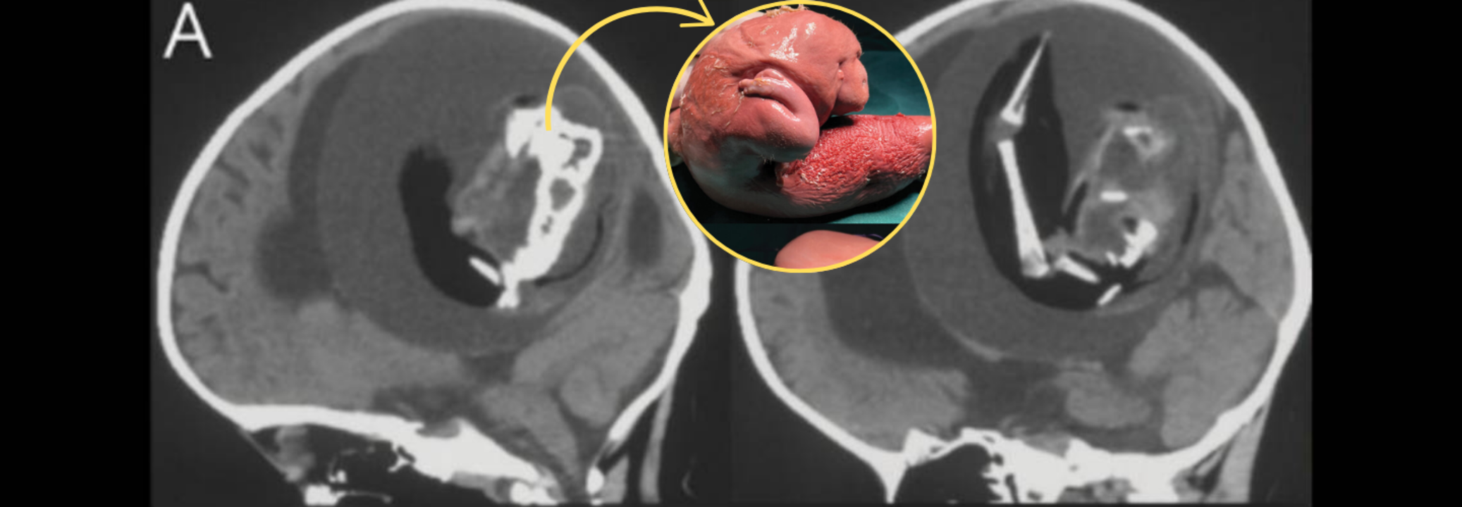
Image Credits: Journal Neurology
In a case that has left the medical community stunned, Chinese doctors successfully extracted a fetus from the brain of a 1-year-old boy. This very rare condition, "fetus in fetu," is when one twin gets engulfed by the other early in fetal development. Most fetus-in-fetu cases are discovered in the abdomen, but this case, where absorption into the brain occurred, is almost unheard of.
The parents of the young girl obtained medical care after they observed that she was developing delayed motor skills and had an abnormally large head circumference. Physicians found an abnormal growth in her skull, which was subsequently diagnosed as a malformed twin. The condition is estimated to happen in only 1 in 500,000 live births, making it a rare occurrence in the history of medicine.
Fetus-in-fetu is a situation where one twin gets trapped within the other in early embryonic development. It happens when identical twins do not fully separate and one embryo gets enclosed within the other. Usually, the engulfed twin does not develop, but the host twin develops normally.
Here, the mass in the infant's brain was a "monochorionic diamniotic twin," which means that the two fetuses initially shared the same placenta but existed in distinct amniotic sacs. In most cases, identical twins completely separate from each other, but in exceptionally rare instances such as this, unseparated blastocysts (early-stage embryonic cells) can result in one twin being imprisoned within the other. As the host embryo grows, it can wrap around the captive twin, incorporating it into its body structure.
Physicians concluded that removal of the parasitic twin by surgery was mandatory to ensure the baby's well-being. Scans of the brain indicated that the deformed fetus had a vertebral column, leg bones, and even the embryonic beginnings of arms and fingers. It also showed spina bifida, where the spinal cord is exposed because of the insufficiency of growth of the tissue around it.
The fetus was about 10 centimeters long and was trapped in a fluid-filled sac inside the child's head. With the unprecedented nature of such a case, medical experts were confronted with an untested challenge of conducting the delicate operation. The operation was completed successfully, excising the fetal mass without any reported complications, although the child's post-operative status has not been made public.
Fetus-in-fetu can be confused with a teratoma, another tumor type which also involves undifferentiated tissue such as hair, teeth, and bone. Fetus-in-fetu differs, however, in that it is the result of a real twin instead of abnormal cellular development. Fetus-in-fetu is identified most commonly based on the formation of a vertebral column and internal structures which imply fetal development.
The fact that the fetal twin was found within the brain indicates that the condition presumably occurred during a developmental stage of neural plate folding. This step is important to the development of the brain and spinal cord. If a parasitic twin were trapped at this time, then it could get integrated into the forming nervous system, as noted in this scenario.
Genome-wide sequencing verified that the fetal mass was genetically equivalent to the surviving twin, further establishing its status as a malformed identical twin instead of a tumor.
Although fetus-in-fetu has been previously reported, cases in which the brain is involved are rare. Perhaps one of the earliest recorded instances occurred in 1982 when a 6-week-old infant underwent surgery to have a parasitic twin removed from inside its skull. That fetus was 14 centimeters long and had formed limbs, a torso, and a head.
The rarity of such cases makes them highly significant for medical research. Understanding how and why fetus-in-fetu occurs can provide insights into early fetal development and the mechanisms that lead to identical twinning. Additionally, these cases highlight the importance of early medical intervention when developmental abnormalities are detected in infants.
These cases underscore the need for prenatal and postnatal medical examinations. Early diagnosis by the use of sophisticated imaging technologies like MRI and CT scans may identify such anomalies early enough to prevent them from causing life-threatening complications.
Though much is yet to be discovered about fetus-in-fetu, this case will remain a vital point of reference for all future medical research. It also makes one more aware of the intricacies of fetal development and the unusual conditions that can develop as a result of anomalies in early cell division.
The excision of a malformed twin from the brain of a 1-year-old is a milestone in pediatric neurosurgery. It highlights the wonders of contemporary medicine and the need for ongoing research to comprehend unusual congenital conditions. With advancing medical science, these findings will make early diagnosis and treatment options more effective for future cases of this nature.